An HCP antibody coverage analysis, in SpotMap 2D
A 45-second loop of the SpotMap 2D workflow: a 2D gel and its matching 2D western blot checked on import, automatic alignment, automatic spot detection on the gel copied to the blot, present or absent calls, antibody coverage overall and per quadrant, then a PDF report signed off in AuditSafe.
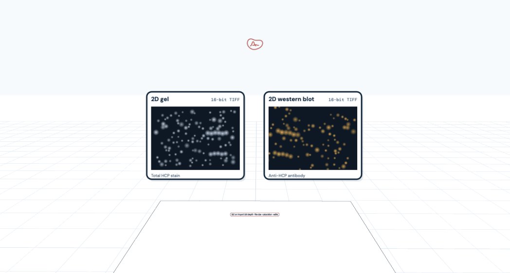
1 / 6 · Import
Gel and blot in, checked on import.
Drag in a 2D gel and its 2D western blot from any scanner. QC flags bit depth, file size, saturation and edits.
HCP antibody coverage by 2D western blot, overall and per quadrant, with automatic alignment and spot detection. A coverage percentage in about 10 minutes.
Start a free trial at totallab.comYour software won’t do what you need? Ask TotalLab.
Import and QC
Drag 2D gel and 2D western blot images straight in from almost any scanner, as TIFF, GEL, MEL, IMG/INF, GSC, 1SC, JPEG, PNG or BMP files. QC on import flags bit depth, file size, saturation and edits. Crop, adjust contrast, flip, rotate or invert inside SpotMap 2D while the TIFF metadata is preserved. Replicate gel and blot pairs can all sit in one experiment file.
Automatic alignment
Pixel-level alignment lays the blot over the gel in seconds and removes the positional variation of 2D gels. Align gel to blot, gel to gel or any combination, and add manual vectors for tricky pairs.
Automatic spot detection
Spots are detected automatically on the gel, which is cleaner than the blot, and the one spot map is copied to every image in the project, so each spot is compared at exactly the same place: 100% matching, with no missing values. Smoothing and peak sensitivity are adjustable, and spots can be added, removed, split or merged.
Present or absent
Every spot is marked present or absent on the blot, one at a time or objectively with filters on area, volume or peak height. Toggle a borderline call and the coverage recalculates immediately. Drug substance spots can be grouped as product spots and kept out of the score.
Coverage score
One coverage percentage for the experiment, by spot number or spot volume, plus a separate score for each quadrant (high and low molecular weight, acidic and basic pI), so a weak corner of the map is visible rather than averaged away. Coverage is the orthogonal check used when qualifying the antibody behind an HCP ELISA (USP <1132>).
Report and sign-off
Build a customizable PDF report with the QC summary, parameters, results and coverage charts. With AuditSafe, SpotMap 2D runs under 21 CFR Part 11 and EU Annex 11 controls, with user access control, activity logging and project sign-off.
“We have had real difficulty matching reactions on blots to the corresponding areas on the gels. […] Once we had the images put through SpotMap software it corresponded to areas that personally I would have never matched, which ended up being the correct areas.”
Craig Pattinson, Research Technician, Proteomics, Aberdeen University
Try SpotMap 2D free on your own gel and blot pairs, with a coverage percentage in about 10 minutes. Recommended by Merck for use with its Auto2D devices. Working under GMP? AuditSafe adds 21 CFR Part 11 and Annex 11 controls.
Start free trial