
2D DIGE vs 2D Gel Electrophoresis

What is 2D DIGE?
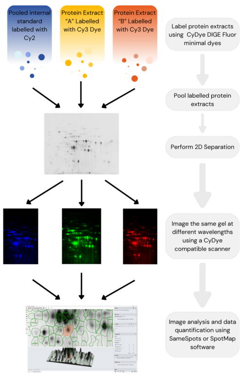
When performing 2D electrophoresis, proteins are separated by their pI (isoelectric point) using isoelectric focusing (IEF) (1st dimension) and then by their molecular weight in SDS-PAGE (2nd dimension).

Figure adapted from Chapter 4, 2-D Fluorescence Difference Gel Electrophoresis (DIGE) in Neuroproteomics, Roberto Diez, Michael Herbstreith, Cristina Osorio, and Oscar Alzate
Two-dimensional difference gel electrophoresis (2D DIGE) is a technical improvement beyond the standard 2D gel electrophoresis protocol. In addition to all the above benefits of 2D gel electrophoresis, 2D DIGE labels different samples with up to 3 fluorescent tags. This allows up to 3 samples to be run on the same gel—saving cost, minimizing gel-to-gel variation, and leading to higher sensitivity/accuracy.
Advantages of 2D DIGE over 2D Gel Electrophoresis:
| 2D DIGE | Standard 2D Electrophoresis | |
| Labelling Method | Fluorescent Dye | Stain |
| Spot Sensitivity | 0.2 ng/spot | Coomassie Blue: 50 ng/spot |
| Silver Staining: 1 ng/spot | ||
| Spyro Ruby: 1 ng/spot | ||
| Number of samples per 2D gel | 3 per gel | 1 per gel |
| Number of in-gel comparisons | 3 | 1 per every 2 gels |
| Spot Resolution | Higher | Lower |
| Reproducibility | Higher | Lower |
- Higher sensitivity: 2D DIGE uses Fluorescent labeling with a sensitivity of 0.2 ng/spot, versus the sensitivity of standard 2D gel electrophoresis with Coomassie blue staining at 100 ng/spot or silver staining at 1 ng/spot. This means you will often detect spots using fluorescent labels that you wouldn’t with standard stains.
- Higher accuracy: The extremely high spot resolution enables greater accuracy when performing spot quantitation. Variances in protein expression as small as +/- 10% can be detected
- Higher reproducibility: Nearly identical data can be obtained using the same sample labeled with different CyDye on the same gel or across different gels, thus eliminating the need to run technical replicates
- Fewer number of gels required: Each 2D DIGE gel can accommodate up to 3 different samples, versus 1 sample on each 2D Gel. For the same number of samples, only 1/3 number of 2D DIGE gels is needed. This is a huge saving in terms of labour and material cost.
How TotalLab’s software can help
Here at TotalLab we have two pieces of software designed specifically for the analysis of 2D electrophoresis gels and blots and both are compatible with experiments performed using 2D DIGE:
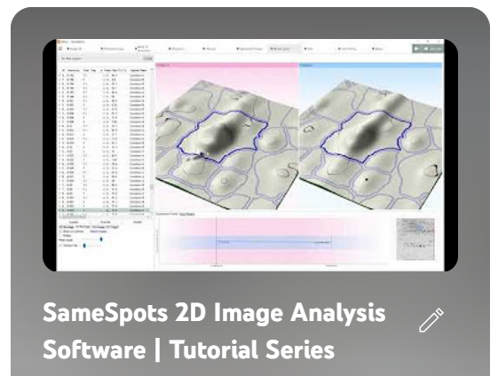
- SameSpots is designed for differential protein expression analysis in large-scale proteomic experiments where multiple different gels need to be compared across multiple, complex experimental conditions (treated vs control, longitudinal studies etc.)
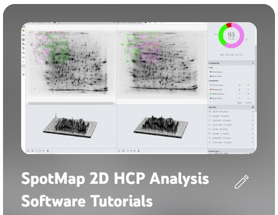
- SpotMap is the only software in the world designed specifically for calculating anti-HCP antibody coverage to validate host cell protein-specific antibodies used in biotherapeutic production to purify drug products. It’s also the only HCP-focused software in the world that is available in a 21-CFR/GMP-compliant version to allow you to comply with FDA regulations surrounding the use of orthogonal techniques to validate anti-HCP antibodies.
You can find more information, including demos and tutorials on our range of 2D image analysis software, here:
 |
 |