2D DIGE vs ELISA vs AAE for HCP Analysis

2D DIGE Western Blot vs Standard 2D vs ELISA vs AAE
What are the differences between the various techniques for determining HCP antibody coverage?
HCP Antibody coverage can be performed by standard 2D Western Blot, ELISA, and AAE. When compared to standard 2D Western Blotting, 2D DIGE Western Blot is capable of:
- High consistency: Perfect alignment of HCP and WB images from the same gel, eliminating any gel-to-gel or gel-to-membrane variations as seen in the standard 2D WB.
- Cost-effective: Since only one gel is needed, it saves the cost of running duplicate gels.
- High accuracy: Fluorescent CyDye labeling allows accurate quantitation of protein and WB spots.
- High sensitivity: Fluorescent CyDye labeling has a higher sensitivity and wider dynamic range than silver staining.
Antibody affinity extraction (AAE), based on antibody immuno-binding with antigen under native conditions, has significant limitations in quantitating antibody coverage:
- False positives: a) direct and indirect associate proteins;
b) antigens present in the HCP antibody;
c) co-purified serum proteins - False negatives: a) associate proteins as a big complex will block antibody binding;
b) some binding HCPs or their degraded fragments cannot be eluted - Not all HCP proteins/fragments can be affinity purified, and the HCP profile is totally changed after AAE
- In addition, AAE still needs be coupled with downstream LC-MS or 2D Western blot, the additional length of the process introduces more variations and inconsistency in data.
| ELISA | Standard 2D Western Blot | 2D DIGE Western Blot | |
|---|---|---|---|
| Number of gels for each Ab | N/A | Two | One |
| Detection Method | Colorimetric | Silver Staining | CyDye Labeling |
| Detect HCP Composition | No | Yes | Yes |
| Detect protein modification | No | Yes | Yes |
| Detect protein degradation | No | Yes | Yes |
| Protein and WB in same gel | No | No | Yes |
| In-gel protein and WB comparison | No | No | Yes |
| Accuracy | Low | Low | High |
| Inaccurate: In-direct reaction of antibodies with the antigen’s associated proteins | Inaccurate: Protein spots counted from the gel, NOT from membrane | Accurate: Protein spots counted directly from the membrane | |
False negative:
|
Error caused by:
|
Accurate:
|
|
| Consistency & Reproducibility | Lower | Lower | Higher |
How TotalLab’s software can help
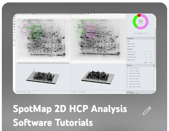
SpotMap is the only software in the world designed specifically for calculating anti-HCP antibody coverage to validate host cell protein-specific antibodies used in biotherapeutic production to purify drug products. It’s also the only HCP-focused software in the world that is available in a 21-CFR/GMP-compliant version to allow you to comply with FDA regulations surrounding the use of orthogonal techniques to validate anti-HCP antibodies.
You can find more information, including demos and tutorials on our SpotMap software here: