
Residual Host Cell Protein Analysis for AAV and Gene Therapy Products: Why the Monoclonal Antibody Playbook Does Not Transfer

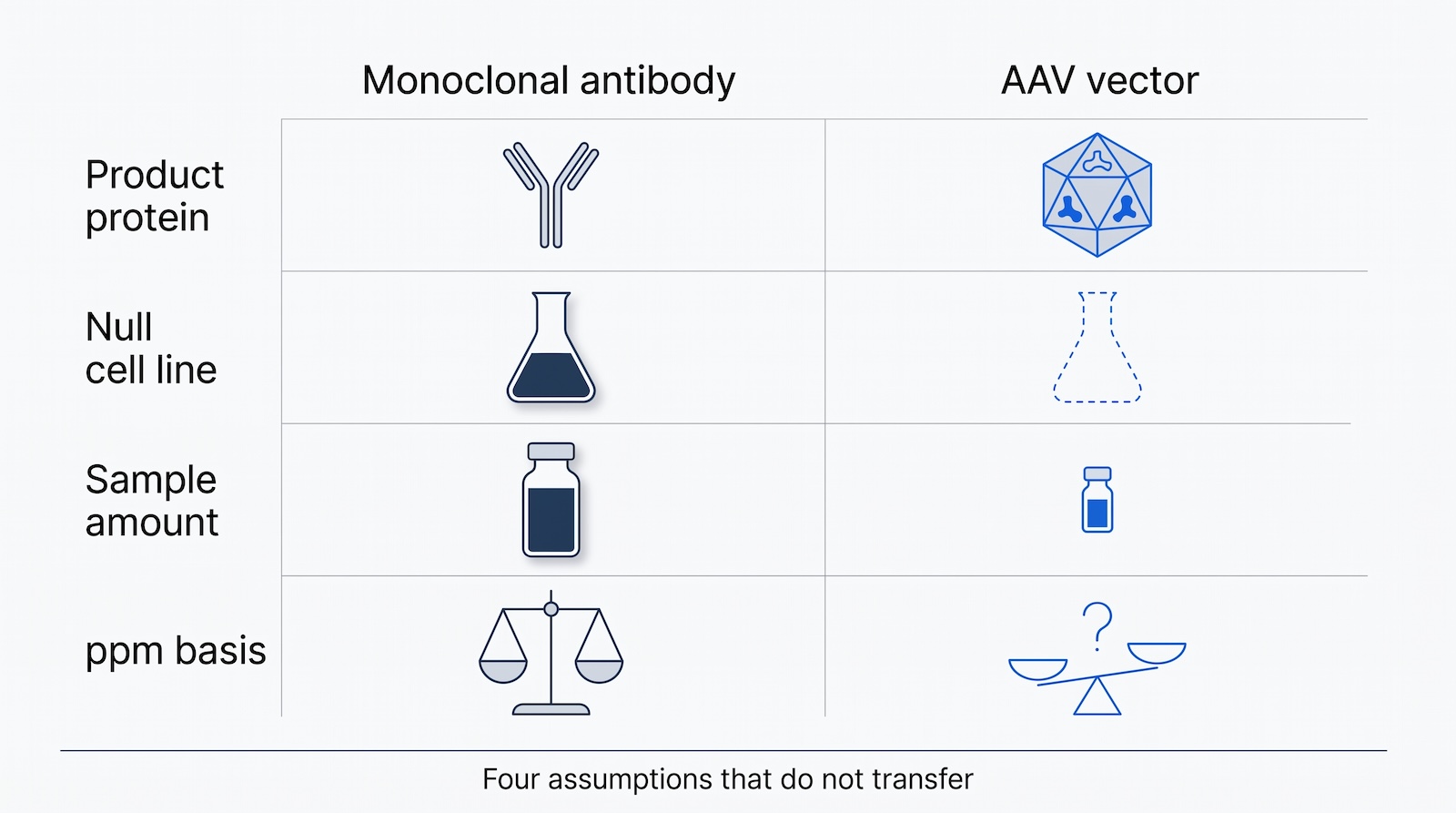
Residual host cell protein (HCP) analysis for adeno-associated virus (AAV) and lentiviral vector (LVV) products is not the monoclonal antibody workflow run on a different cell line. The product protein is a 60-subunit capsid rather than one defined sequence, a dose carries micrograms rather than hundreds of milligrams of it, the production cells are transiently transfected so no honest null lysate exists, and much of the protein burden comes from plasmids and helper virus rather than from the host genome. The reporting denominator is still unsettled. This page sets out what changes, what FDA and EMA actually ask for, why the two USP chapters point in opposite directions on your modality, and how to build an LC-MS method that works on very little material.
Key facts
- Host cell protein (HCP) analysis for adeno-associated virus (AAV) and lentiviral vector (LVV) products is not the monoclonal antibody (mAb) workflow with a different cell line. The product protein, the denominator, the reagents and the impurity classes all change.
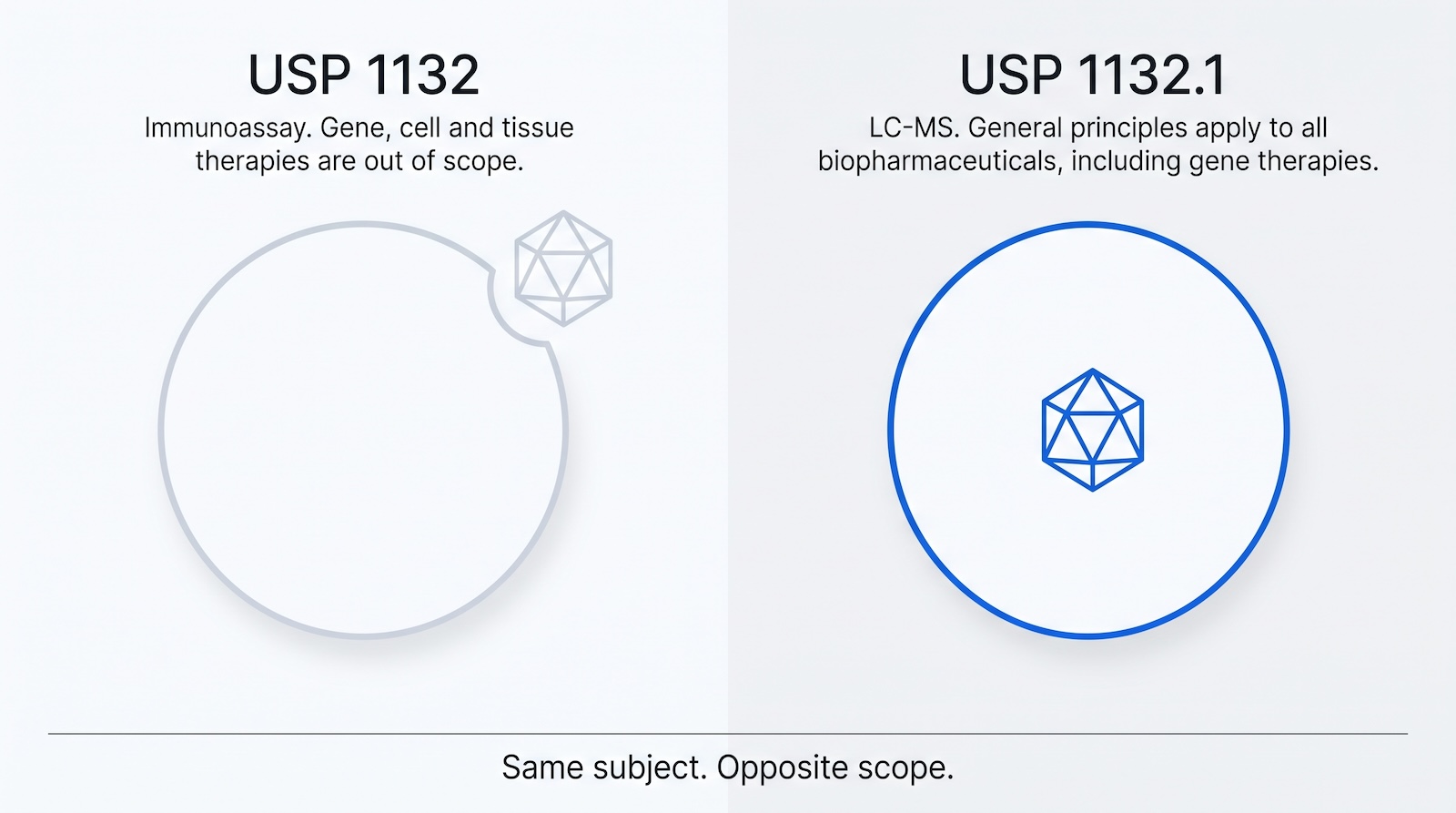
- The two pharmacopeial chapters point in opposite directions, and that asymmetry is the argument for LC-MS. USP General Chapter <1132>, the immunoassay chapter, scopes gene therapy out in its own words: “This chapter focuses on HCP immunoassays for recombinant therapeutic products. It does not address products such as vaccines or gene-, cell-, or tissue-based therapies, although the general principles discussed may apply to the measurement of HCPs in these products.” USP posts the USP 39 and NF 34 version of that chapter, official 1 May 2016, as a free full-text PDF, so you can check that sentence yourself.
- USP General Chapter <1132.1>, the LC-MS chapter, does the opposite. Its Introduction and Scope (Section 1) states that although the chapter focuses on recombinant proteins, the general principles it sets out apply to all types of biopharmaceuticals, and it names vaccines, gene therapies, cellular and tissue based products and biocatalysis products among them. The immunoassay chapter excludes your modality. The mass spectrometry chapter includes it by name.
- One AAV capsid weighs about 3.8 megadaltons, so a 1.5 x 10<sup>11</sup> vector genome subretinal dose carries roughly one microgram of capsid protein, five orders of magnitude less than a 100 mg antibody dose.
- The FDA’s 2020 gene therapy CMC guidance recommends testing for process-related impurities including “residual cell substrate proteins” and “helper virus contaminants (i.e., infectious virus, viral DNA, viral proteins)”. The EMA guideline names “host cell protein (including helper virus protein)”.
- There is still no agreed convention for the reporting unit in a viral vector product. Published work uses ng per microgram of total HCP, ng/mL and mass per vector genome. The method has a pharmacopeial reference in USP <1132.1>; the denominator does not.
Why AAV and lentiviral vectors are different from monoclonal antibodies
They differ in the four things an HCP assay depends on: what the product protein is, how much of it exists, what the host cell is, and what else the process puts in the vial. A monoclonal antibody is a single, well-defined, highly abundant polypeptide pair from a stable Chinese hamster ovary (CHO) line. An AAV vector is a 60-subunit shell built from VP1, VP2 and VP3, made in transiently transfected human embryonic kidney 293 (HEK293) cells or in Sf9 insect cells infected with recombinant baculovirus, and present at microgram quantities.
Take the mass first. Wörner and colleagues measured AAV capsids by charge detection mass spectrometry and describe “a non-enveloped T = 1 icosahedral capsid of ~3.8 megaDalton”, about 6.3 x 10⁻¹⁸ g per capsid. LUXTURNA, an approved AAV2 product, is dosed at “1.5 x 10¹¹ vector genomes (vg), administered by subretinal injection in a total volume of 0.3 mL”. At one capsid per vector genome that dose carries roughly 0.95 micrograms of capsid protein, against 100 milligrams for a 100 mg antibody dose. Do not over-generalize it: ZOLGENSMA is dosed at “1.1 × 10¹⁴ vector genomes per kilogram (vg/kg) of body weight”, so a 5 kg infant receives about 3.5 mg, and empty capsids add more in both cases. Product protein mass per dose spans microgram to low milligram, and the low end is where conventional HCP analytics break.
AAV and LVV vs monoclonal antibody: what changes for HCP analysis
| Attribute | Monoclonal antibody | AAV vector | Lentiviral vector |
|---|---|---|---|
| Expression system | Stable CHO line, fed-batch or perfusion | Transient transfection of HEK293, or Sf9 with recombinant baculovirus | Transient transfection of HEK293T, or stable producer line |
| Product protein definition | One heavy chain plus one light chain, single defined sequence | Three capsid proteins VP1, VP2 and VP3 in variable stoichiometry across capsids | Gag-derived structural proteins (MA, CA, p2), plus protease, reverse transcriptase, integrase and envelope glycoprotein |
| Total protein mass per dose | Tens to hundreds of milligrams | Roughly 1 microgram (subretinal AAV2, 1.5 x 10^11 vg) to a few milligrams (systemic AAV9 in an infant) | Typically sub-milligram; dose is usually expressed in transducing units, not mass |
| Typical sample amount available for LC-MS | Milligrams; 100 microgram digests are routine | Micrograms; published methods use 2 to 50 micrograms total protein | Micrograms; often less, and material is precious |
| ELISA reagent availability | Mature platform and process-specific CHO kits, decades of use | Generic HEK293 kits exist; no widely used Sf9 gene therapy platform kit | Generic HEK293 kits; producer-line-specific reagents rare |
| Null cell line for the ELISA standard | Untransfected parental CHO is a good model of the production cell | Untransfected HEK293 is not the transfected, AAV-producing cell; the proteome shifts on transfection | Same problem, plus the vector particle carries host proteins by design |
| Denominator for ppm reporting | ng HCP per mg product protein, with product protein by A280 or amino acid analysis | Unsettled. Capsid protein mass, total protein, vector genomes and capsid particles are all used | Unsettled. Often per mL or per transducing unit |
| Dominant interfering protein | The antibody itself | VP1, VP2 and VP3 capsid protein | Gag-derived capsid protein; Johnson et al. reported CA sequence coverage of 57% to 79% |
| Additional process-derived proteins | Few; protein A ligand leachate is the main one | Rep and Cap, adenoviral helper proteins, baculovirus proteins in Sf9 processes | Packaging and envelope plasmid products, including VSV-G |
| Pharmacopeial framework | USP 1132 for immunoassay, USP 1132.1 for LC-MS | USP 1132 states it does not address gene-based therapies. USP 1132.1 states its general principles apply to all types of biopharmaceuticals and names gene therapies, so the LC-MS chapter is the one to work from | Same |
| Serotype or pseudotype dependence | Not applicable | Real. Leibiger et al. found greater overlap in conserved HCPs between AAV8 and AAV9 than with AAV2 | Real. Johnson et al. found fewer host protein species in RDpro-pseudotyped vectors than in VSV-G-pseudotyped vectors |
What this does to ppm reporting
HCP ppm means nanograms of host cell protein per milligram of product protein. For an antibody that is unambiguous, and the arithmetic and its failure modes are covered on how HCP ppm is calculated from LC-MS data. For a viral vector the denominator is the problem, and four things go wrong at once. Absorbance at 280 nm cannot be converted cleanly to capsid protein mass, because the packaged DNA genome absorbs strongly in the same region. Total protein by a colorimetric assay includes the host cell proteins themselves. Vector genome titer is not a protein mass, and it diverges from capsid protein mass as the empty capsid fraction changes. And capsid protein mass per particle is not constant.
So a ppm figure is not interpretable unless the report states the denominator. Leibiger and colleagues normalized individual HCPs to ng per microgram of total HCP; other method papers report ng/mL. Report the numerator in absolute mass, state the denominator, and give the vector genome titer alongside.
The product protein is also not one protein: VP1, VP2 and VP3 share C-terminal sequence and differ at the N-terminus, and the nominal 1:1:10 stoichiometry is an average rather than a fact about any particle, so there is no single sequence to divide by.
The host cell differs too. Most clinical AAV comes from transient triple transfection of HEK293 cells, which introduces Rep and Cap proteins and adenoviral helper proteins encoded by plasmids rather than by the host genome. Those are process-related impurities, but not host cell proteins in the sense an HCP ELISA or an HCP database means. Transfection also reshapes the host proteome: Strasser and colleagues compared transfected and untransfected HEK293 cells producing AAV5 and reported 530 proteins significantly differentially expressed in cell pellets and 1,275 in supernatants. The cells that make your product are not the cells in your null lysate. Sf9 production changes the population again, to insect proteins plus recombinant baculovirus to clear; Smith and colleagues found 113 HEK293-derived HCPs in an AAV2 sample and 102 Sf9-derived proteins in an AAV8 sample.

Why HCP ELISA is especially weak for viral vector products
Because the three things it depends on are all weaker here: the immunogen does not match the production cells, coverage is unmeasured against your process, and much of the process-related protein burden is not host cell protein at all. USP General Chapter <1132>, the reference text for HCP immunoassay design, states plainly: “This chapter focuses on HCP immunoassays for recombinant therapeutic products. It does not address products such as vaccines or gene-, cell-, or tissue-based therapies, although the general principles discussed may apply to the measurement of HCPs in these products.”
That sentence is worth reading twice, and it is worth checking rather than taking on trust. USP posts the USP 39 and NF 34 version, official 1 May 2016, as a free PDF, so the scope statement is one click away. Note the version when you cite it; no Notice of Intent to Revise has been published for <1132> since it became official. The sentence does three things at once. It is not a prohibition, and the chapter says so itself: the general principles “may apply”. It is an explicit statement that the chapter’s specific requirements, on immunogen preparation, null cell lines, coverage assessment and acceptance criteria, were written with a stable recombinant protein process in mind and were not validated against a transiently transfected viral vector process. And it puts the burden of justification on you rather than on a reviewer, because a sponsor who borrows the structure of <1132> for an AAV product is applying a document that names their modality as out of scope. Say so in the method description, explain which principles you adopted and why, and the borrowing becomes a documented choice rather than an unexamined assumption.
The null cell line requirement is where it bites. USP <1132> describes establishing “a null cell that does not express the product gene”, using parental cells or cells transfected with the vector minus the product coding gene. For a stable CHO line that is a good model. For transient AAV production it is not, because transfection reshapes the proteome by hundreds to over a thousand proteins. An antibody raised against untransfected HEK293 lysate is raised against a different population from the one in your harvest.
Generic kits exist and are used. Cygnus Technologies says the antibodies in its HEK 293 HCP ELISA Kit, 3G were “generated against and affinity-purified using a mild lysate of HEK 293 cells”, with “broad coverage of ~90% to HEK 293 HCPs as determined by Antibody Affinity Extraction with 2D PAGE/Silver Stain and LC-MS”. That is coverage against a HEK293 lysate, not against the residual population surviving your capture and polishing. Coverage is a property of the antibody-plus-process pair, as set out in why you need to know your HCP antibody coverage.
An ELISA also cannot see what it was not raised against. Rep and Cap, adenoviral helper proteins and baculovirus proteins are absent from a null HEK293 or Sf9 lysate by construction, so a generic kit returns nothing about them even though FDA and EMA name them explicitly. Smith and colleagues conclude that ELISA “has limited specificity and has been shown to underestimate the overall HCP content of AAV produced in mammalian cells”. Immunoassay and mass spectrometry disagree for CHO products too, for reasons covered in why ELISA and LC-MS HCP numbers disagree, but here the mismatch is larger.
The immunoassay chapter excludes you. The LC-MS chapter names you.
USP General Chapter <1132.1>, the LC-MS companion chapter published in USP-NF 2025 Issue 1 and official from 1 May 2025, does not exclude gene therapy. Its Introduction and Scope says the chapter focuses on recombinant proteins but that the general principles it sets out are “applicable to all types of biopharmaceuticals” (USP <1132.1>, Section 1), and it names the categories it means: vaccines, gene therapies, cellular and tissue based products, and biocatalysis products. Gene therapy is on that list by name. That is the opposite of what <1132> says, and it is the most useful pharmacopeial fact available to a gene therapy CMC team.
Put the two chapters side by side and the pharmacopeia has already made the technical argument for you. The immunoassay chapter, the one that describes the null cell line, the polyclonal immunogen and the coverage assessment, says it does not address your modality. The mass spectrometry chapter, the one that describes bottom-up digestion, database searching, spiked standards and system suitability, says its principles apply to all types of biopharmaceuticals and names gene therapies among them. That asymmetry is not an accident. An immunoassay depends on a reagent raised against a specific cell population, which is exactly the thing a transiently transfected viral vector process does not give you. A mass spectrometry method depends on sequences and standards, which transfer across modalities in a way antibodies do not. Read together, the two chapters are an argument for building the LC-MS method rather than stretching an ELISA designed for a different class of product.
The practical consequence for a gene therapy program is that you are not working in a pharmacopeial vacuum. You cannot claim compliance with either chapter, because both are informational chapters numbered above 1000 and neither carries mandatory requirements. What you can do is say in your method description that the method follows the principles of USP <1132.1>, name the sections you followed, and note that the chapter states those general principles are applicable to all types of biopharmaceuticals including gene therapies. That is a stronger position than borrowing the structure of a chapter that names your modality as out of scope, and it is available today.
Two caveats keep it honest. The chapter is written around recombinant protein examples, so the specifics, the reporting units above all, still need adapting and justifying for a vector product. And all of <1132.1> sits behind a USP-NF subscription, so a reader without a license cannot check the scope statement the way they can check the free <1132> PDF. Cite the section number and the DOI, and say access is by subscription.

What regulators actually expect
Process-related impurity control is a named expectation in both the FDA and EMA frameworks for gene therapy, but neither sets a numeric HCP limit and neither prescribes a method. The FDA guidance “Chemistry, Manufacturing, and Control (CMC) Information for Human Gene Therapy Investigational New Drug Applications (INDs)”, issued January 2020 by CBER and still in force, states: “We recommend testing for process-related impurities. These include, but are not limited to, residual cell substrate proteins, extraneous nucleic acid sequences, helper virus contaminants (i.e., infectious virus, viral DNA, viral proteins), and reagents used during manufacture”. Note what it does not do. It does not name an assay, set a limit, or require mass spectrometry.
The EMA equivalent is the Guideline on the quality, non-clinical and clinical aspects of gene therapy medicinal products (EMA/CAT/80183/2014, effective 22 March 2018). Its drug substance specification section states that “tests should be developed and relevant (upper) limits set to monitor the residual levels of contaminants of cellular origin, e.g. host cell protein (including helper virus protein) or DNA from the bacterial or packaging cell line”. The parenthesis matters: it folds helper virus protein into the same expectation as host cell protein.
FDA published “Flexible Requirements for Cell and Gene Therapies to Advance Innovation” in January 2026, but it covers release specifications and process validation, not impurity expectations. So you are expected to control residual protein, justify your method and set limits you can defend, not to hit a published number. The pharmacopeial position is better than it is usually reported. USP <1132>, the immunoassay chapter, says it does not address gene-, cell- or tissue-based therapies. USP <1132.1>, the LC-MS chapter, says its general principles apply to all types of biopharmaceuticals and names gene therapies among them. Neither is mandatory, because both are informational chapters numbered above 1000, so following <1132.1> is not a compliance shortcut. It is something better in a filing: a named, current, independent reference for the method you built, that does not exclude your modality. If you follow its quantitation methods, Sections 5.1.1, 5.1.2 and 5.1.3, name the section you used and say why, as described in running USP <1132.1> Methods A, B and C.
The analytical approach: LC-MS on very little material
LC-MS is the practical route because it needs no matched antibody reagent, it identifies proteins rather than returning one immunoreactive total, and it detects plasmid- and helper-derived proteins in the same run as host cell proteins. Bracewell and colleagues note that the approach “is being established, although such information remains limited”.
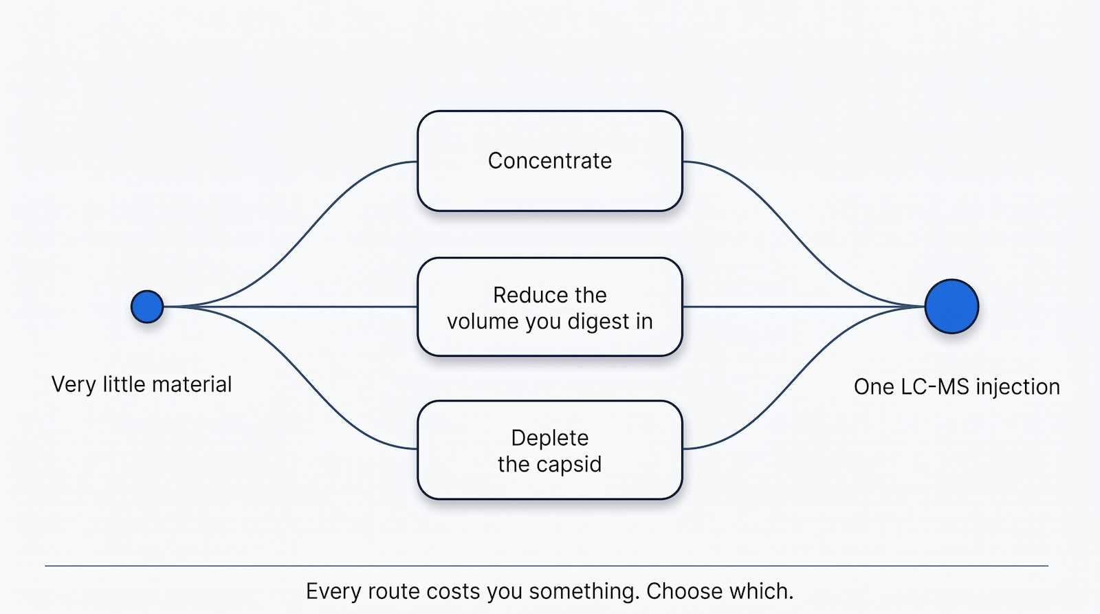
Sample amount is the first constraint. Published AAV HCP methods run on 2 to 50 µg of total protein, one to two orders of magnitude below routine mAb practice. Smith and colleagues used SP3, the single-pot solid-phase-enhanced sample preparation method of Hughes and colleagues, processing 10 µg per digestion on magnetic beads. SP3 suits this work because it gives “virtually lossless and unbiased recovery of proteins independent of input quantity” and tolerates detergents, chaotropes and salts that would otherwise need a lossy removal step. AAV formulations commonly contain poloxamer surfactants.
Capsid protein dominance is the second. This is the same problem an antibody creates for its own HCP assay, and Lodge and colleagues state it directly: “highly abundant biotherapeutics hamper the detection of low-level HCPs”. They adapted native digestion, developed for intact antibodies, to AAV, and found it “more effective than applying denaturing conditions to extract the HCPs associated with different AAV serotypes”. Others exploit the capsid’s resistance instead: Leibiger and colleagues chose trypsin because it “has been shown to have a limited ability to digest AAV viral capsid proteins, and is therefore recommended for protein digestion of rAAV samples to increase relative HCP signal intensity compared to viral capsid proteins”. Enrichment is a third option: Zhang, Xiao and Li applied ProteoMiner beads to AAV products.
Acquisition mode is the third. Data-independent acquisition (DIA) is acquisition in which the instrument fragments all precursor ions in defined mass windows rather than selecting individual precursors, so quantitation does not depend on a precursor being picked in that run. Leibiger, Min and Lee compared four SWATH-MS workflows, SWATH being a DIA implementation, across rAAV2, rAAV5, rAAV8 and rAAV9 produced in HEK293 and purified by AAVX affinity chromatography. Their best configuration, an in silico spectral library processed in DIA-NN on a ZenoTOF 7600, gave “a 78% increase in HCP identifications, 80% reduction in sample requirement, and 70% reduction in instrument runtime” against their earlier DDA-library Skyline workflow, with “median HCP quantitation CV below 10%”. Total sample requirement fell from 30 µg to 6 µg. Building a library by data-dependent acquisition consumes sample you do not have; a predicted library removes that cost. Software that takes a user-supplied FASTA and a predicted library and applies identical rules to every run is what makes this repeatable under quality control, and TotalLab’s SpotMap MS is one such DIA-based package. The general comparison is in DIA vs DDA for HCP analysis.
Serotype-dependent impurity profiles are the fourth. Leibiger and colleagues quantified residual HCPs across four serotypes after identical affinity capture, identified “2,746 unique protein species derived from the producing HEK293 cell line” in at least one sample, and found heat shock 70 kDa protein 1B (HSPA1B), Hsc70-interacting protein (ST13) and heat shock protein HSP 90-beta (HSP90AB1) most persistent. AAV8 and AAV9 shared more conserved HCPs with each other than either did with AAV2, so a clearance profile does not transfer between serotypes. Recurring named impurities are the candidates for targeted methods, on the logic set out for high-risk host cell proteins.
Sample preparation considerations for low-input viral vector samples
| Consideration | Why it matters for viral vectors | Practical option | Watch for |
|---|---|---|---|
| Input amount | Published AAV methods run at 2 to 50 micrograms total protein; mAb practice assumes 100 micrograms | SP3 on magnetic beads; Smith et al. used 10 micrograms per digestion | Adsorptive loss to tubes and tips dominates at low input; use low-bind consumables and minimize transfers |
| Surfactant in formulation | Poloxamer 188 and Pluronic F-68 are common in AAV formulations and suppress ionization | SP3 tolerates detergents and washes them away; bead-based enrichment also removes Pluronic F-68 | Incomplete removal shows up as suppressed signal across the whole run, not as a single bad peptide |
| Excipient and salt load | Small dose volumes mean excipient is a large fraction of the sample | Choose a prep that binds protein and washes, rather than one that relies on dilution | Dilution to reduce excipient also dilutes the HCPs below detection |
| Digestion strategy | Capsid protein dominates the sample | Native digestion preferentially releases HCP peptides from intact particles; denaturing digestion gives fuller capsid coverage | The two answer different questions. Choose one per method and do not mix them within a comparison |
| Enzyme choice | Trypsin digests AAV capsid proteins poorly, which raises relative HCP signal | Trypsin is the default in published rAAV HCP methods for this reason | The same property makes trypsin a poor choice if you also want accurate VP quantitation in that run |
| Capsid or product depletion | Removing the abundant protein raises HCP depth | ProteoMiner or equivalent equalization beads | Any enrichment step changes recovery protein by protein, so quantitation becomes relative unless you qualify it |
| Spectral library | Building a DDA library consumes sample you may not have | In silico predicted library; Leibiger et al. report an 80% reduction in sample requirement with this route | Predicted libraries depend entirely on the FASTA. A wrong or incomplete FASTA silently limits what can be found |
| FASTA content | Plasmid- and helper-derived proteins are not in a host proteome database | Add Rep, Cap, adenoviral helper and, for Sf9 processes, baculovirus sequences to the search space | Adding sequences expands the search space and affects false discovery rate control; record the exact database version |
| Spiked standards | Absolute quantitation needs a reference of known amount | A spiked protein or stable isotope labeled peptide, as in USP 1132.1 Sections 5.1.2 and 5.1.3 (Methods B and C) | The spike must experience the same prep; at low input a protein spike can dominate the digest |
| Replication | Low-input preps are more variable | Triplicate injections at minimum; Leibiger et al. report median CV below 10% for their best method | A CV computed across injections is not the same as a CV across independent preparations |

VP1:VP2:VP3 ratio and capsid quantitation, briefly
Capsid protein quantitation is an adjacent measurement on much of the same instrumentation. Wörner and colleagues describe the nominal VP1:VP2:VP3 ratio as “estimated to be in a ratio of 1:1:10 based on gel densitometry and mass spectrometry studies”, then show that real preparations depart from it: assembly is stochastic, and their measured empty capsid mass distribution “spans more than 300 kDa of very densely populated masses between 3.6 and 3.9 MDa”. Capsid stoichiometry is a distribution, not a number.
Kontogiannis and colleagues published an LC-MS method in 2025 that quantifies VP1, VP2 and VP3 in rAAV9 and profiles host cell proteins in the same workflow, the clearest case for treating the two readouts as one method: one prep, one injection, two reportable attributes. Empty and full capsid ratio is a third attribute and a separate method; FDA classifies “empty capsid particles” as a product-related impurity, distinct from the process-related impurities above, and it is not an HCP measurement.
Lentiviral vectors: two impurity populations, one assay
Lentiviral vectors add a complication. The particle is enveloped and acquires host membrane and cytosolic proteins as it buds, so some host proteins are structurally part of the product. Separating “residual HCP” from “particle-associated host protein” is a judgment call the field has not settled.
Johnson and colleagues analyzed size-exclusion-purified lentiviral vectors by LC-MS/MS and identified 93 different cellular proteins, with beta-actin, heat shock cognate 71, clathrin heavy chain 1, ALIX and cyclophilin A appearing consistently. Pseudotype mattered: “transiently produced VSV-G-pseudotyped vectors contain considerably more host protein species compared to stably produced RDpro-pseudotyped vectors”, which the authors attribute in part to co-purifying VSV-G vesicles. Change the envelope, or move from transient to stable production, and the impurity profile changes with it.
The same run detected the vector’s own proteins: “Peptides of viral proteins, including HIV Gag protein domains (matrix (MA), capsid (CA) and p2), protease (PR), reverse transcriptase (RT), integrase (IN) and VSV-G Env were detected in all 3 sets of purified vectors”. That is the argument for one LC-MS method rather than a panel. A p24 ELISA for particle content, a HEK293 HCP ELISA for host protein and nothing for packaging plasmid products gives three disconnected numbers and a gap; one characterized proteomic method gives an inventory.
A practical workflow, and where it is hard
- Decide what you are counting before you start. Host cell proteins only, or host cell proteins plus plasmid- and helper-derived proteins? It determines the FASTA, the reporting units and whether the result compares to anything else, and it causes most of the later arguments.
- Build the search space deliberately. Host proteome (human for HEK293, Spodoptera frugiperda for Sf9), plus Rep and Cap for your serotype, your adenoviral helper genes, baculovirus sequences for insect cell processes, and common contaminants. Record the database version. A protein absent from the FASTA cannot be found, and nothing in the output will say so.
- Budget the sample. Work out how many micrograms of total protein you can spare, then pick a method that fits. Under about 10 µg, a DDA-built spectral library is out of reach.
- Choose a digestion strategy and commit. Native digestion for maximum HCP depth against an intact capsid, denaturing digestion if you also need capsid sequence coverage. Trypsin’s poor digestion of capsid proteins helps HCP work and hurts VP quantitation.
- Qualify the preparation at the input amount you will use. Recovery, carryover and adsorptive loss behave differently at 5 µg than at 100 µg. Data generated at 100 µg does not qualify a 5 µg method.
- Acquire in DIA with a fixed processing pipeline. Lock the library, software version, false discovery rate threshold and inference rules before generating release data. Reprocessing old data with a new pipeline changes the answer.
- Report absolute mass plus an explicit denominator. Give the mass of each protein, the total, the denominator and how you measured it, and the vector genome titer. Never report a bare ppm figure.
- Trend across the process, not just at release. Clearance across capture and polish is the most useful output during development, and it supports a phase-appropriate specification.
The hard parts are real. Sample is scarce and every development run consumes it. There is no reference material with certified HCP content for a viral vector, so accuracy is assessed against spikes rather than truth. And while USP <1132.1> gives the method a pharmacopeial reference that covers your modality, it does not settle the reporting denominator for a capsid product, so two competent labs can still make different, equally defensible choices and produce numbers that do not compare.
Bringing this in-house
Gene therapy programs run on small sample volumes and short timelines, a poor fit for shipping material to a service lab and waiting. Running the analysis yourself keeps the sample in the building and the method under your control. SpotMap MS is TotalLab’s DIA or DDA-based LC-MS HCP analysis software, and it pairs with AuditSafe for 21 CFR Part 11 and GMP compliance. If you are weighing build versus buy, start with bringing HCP analysis in-house, or request a free trial and run your own AAV data through it.
Frequently asked questions
Do I need HCP testing for AAV gene therapy products, and does any pharmacopeial chapter cover it?
Yes to the first, and yes to the second more than most people think. The FDA’s 2020 gene therapy CMC guidance recommends testing for process-related impurities including residual cell substrate proteins and helper virus proteins, and the EMA guideline requires tests and upper limits for host cell protein including helper virus protein. Neither sets a numeric limit or names a method. On the pharmacopeial side the two chapters diverge: USP <1132>, on immunoassay, states it does not address gene-based therapies, while USP <1132.1>, on LC-MS, states its general principles apply to all types of biopharmaceuticals and names gene therapies. Work from <1132.1>.
Can I use a CHO HCP ELISA for a HEK293 product?
No. A CHO HCP ELISA uses polyclonal antibodies raised against Chinese hamster ovary cell lysate. Human embryonic kidney 293 proteins are different proteins. The assay would report near-zero regardless of the true impurity level. Use a HEK293 reagent, and understand that even a HEK293 kit was raised against untransfected lysate rather than the transfected, vector-producing cells in your process.
How do you report HCP ppm for AAV?
There is no settled convention, so state your basis explicitly. Report the absolute mass of each protein and the total, name the denominator (capsid protein mass, total protein, or per vector genome) and the assay used to measure it, and give the vector genome titer alongside. Both USP chapters define ng/mg as nanograms of HCP per milligram of product protein, and USP <1132.1> says its general principles apply to all types of biopharmaceuticals, but “product protein” is unambiguous for an antibody and is not for a 60-subunit capsid. Adapt the unit and justify the adaptation rather than assuming it transfers.
Is there a null cell line for an AAV HCP ELISA?
Not a good one. USP <1132> describes using parental cells or cells transfected without the product gene as the null. For transient AAV production, transfection itself changes the host proteome substantially: Strasser and colleagues found 530 proteins significantly differentially expressed in transfected HEK293 pellets and 1,275 in supernatants. An untransfected lysate is a different protein population from the one in your harvest.
Does the AAV serotype change the residual HCP profile?
Yes. Leibiger and colleagues profiled rAAV2, rAAV5, rAAV8 and rAAV9 produced in HEK293 and purified identically by AAVX affinity chromatography, and found serotype-dependent overlap: AAV8 and AAV9 shared more conserved HCPs with each other than either did with AAV2. Do not assume clearance data from one serotype transfers to another on the same platform.
How much sample do I need for LC-MS HCP analysis of a viral vector?
Published methods run on 2 to 50 micrograms of total protein. Smith and colleagues used 10 micrograms per SP3 digestion. Leibiger and colleagues reduced their requirement from 30 micrograms to 6 micrograms total by switching to a predicted spectral library, DIA-NN processing and a more sensitive instrument. Qualify recovery at the input amount you will actually use, not at a higher one.
Do plasmid and helper virus proteins count as host cell proteins?
Strictly, no. Rep, Cap, adenoviral helper and baculovirus proteins are encoded by plasmids or a helper virus, not by the host cell genome, so they sit outside a host proteome database and outside a null-lysate-derived ELISA reagent. Both FDA and EMA name them as process-related impurities to control, and the EMA text specifically includes helper virus protein in the same sentence as host cell protein.
Can one LC-MS method measure both capsid proteins and residual HCPs?
In principle yes, and Kontogiannis and colleagues published a method in 2025 that quantifies VP1, VP2 and VP3 in rAAV9 and profiles host cell proteins in the same workflow. In practice there is a tension: trypsin’s poor digestion of AAV capsid proteins is what raises relative HCP signal, and it is also what makes accurate VP quantitation harder in the same run. Expect a compromise or two methods.
References
- Leibiger, T.M., Min, L., Lee, K.H. “A comparison of SWATH-MS methods for measurement of residual host cell proteins in adeno-associated virus preparations.” Frontiers in Bioengineering and Biotechnology, 2 May 2025. DOI 10.3389/fbioe.2025.1579098. https://www.frontiersin.org/journals/bioengineering-and-biotechnology/articles/10.3389/fbioe.2025.1579098/full
- Leibiger, T.M., Min, L., Lee, K.H. “Quantitative proteomic analysis of residual host cell protein retention across adeno-associated virus affinity chromatography.” Molecular Therapy: Methods & Clinical Development, 2024, 32(4):101383. DOI 10.1016/j.omtm.2024.101383. https://pmc.ncbi.nlm.nih.gov/articles/PMC11650319/
- Smith, J., Strasser, L., Guapo, F., Milian, S.G., Snyder, R.O., Bones, J. “SP3-based host cell protein monitoring in AAV-based gene therapy products using LC-MS/MS.” European Journal of Pharmaceutics and Biopharmaceutics, 2023, 189:276-280. DOI 10.1016/j.ejpb.2023.06.019. https://concept-nibrt.ie/wp-content/uploads/2023/11/SP3-based-host-cell-protein-2023.pdf
- Lodge, J., Huang, L., Lian, Z., Qian, J., Tian, Y. “Native Digestion and Shotgun Proteomics for Host Cell Protein Profiling of Adeno-Associated Viruses.” Analytical Chemistry, 2024. DOI 10.1021/acs.analchem.4c00893. PMID 39376079. https://pubs.acs.org/doi/10.1021/acs.analchem.4c00893
- Huo, S., Nie, S., Cong, Y., Wang, S., Li, N. “In-Depth Host Cell Protein Analysis and Viral Protein Impurity Monitoring in Adeno-Associated Virus-Based Gene Therapy Products Using Optimized Wide Window Data-Dependent Acquisition Method.” Analytical Chemistry, 2024. DOI 10.1021/acs.analchem.4c02400. PMID 39263887. https://pubs.acs.org/doi/10.1021/acs.analchem.4c02400
- Zhang, S., Xiao, H., Li, N. “Analysis of Host Cell Proteins in AAV Products with ProteoMiner Protein Enrichment Technology.” Analytical Chemistry, 2024. DOI 10.1021/acs.analchem.3c03884. PMID 38262068. https://pubs.acs.org/doi/10.1021/acs.analchem.3c03884
- Kontogiannis, T., McElroy, C., Quaglia, M., Whale, A.S., Foy, C., Braybrook, J., Smales, C.M. “Development of LC-MS methods for AAV capsid protein quantification and host cell protein profiling.” Molecular Therapy: Methods & Clinical Development, 2025, 33(3):101562. DOI 10.1016/j.omtm.2025.101562. https://kar.kent.ac.uk/111135/
- Johnson, S., Wheeler, J.X., Thorpe, R., Collins, M., Takeuchi, Y., Zhao, Y. “Mass spectrometry analysis reveals differences in the host cell protein species found in pseudotyped lentiviral vectors.” Biologicals, 2018, 52:59-66. DOI 10.1016/j.biologicals.2017.12.005. https://discovery-pp.ucl.ac.uk/id/eprint/10042724/8/Takeuchi_1-s2.0-S1045105617301537-main.pdf
- Bracewell, D.G., Smith, V., Delahaye, M., Smales, C.M. “Analytics of host cell proteins (HCPs): lessons from biopharmaceutical mAb analysis for Gene therapy products.” Current Opinion in Biotechnology, 2021, 71:98-104. DOI 10.1016/j.copbio.2021.06.026. https://kar.kent.ac.uk/89635/
- Strasser, L., Boi, S., Guapo, F., Donohue, N., Barron, N., Rainbow-Fletcher, A., Bones, J. “Proteomic Landscape of Adeno-Associated Virus (AAV)-Producing HEK293 Cells.” International Journal of Molecular Sciences, 2021, 22(21):11499. DOI 10.3390/ijms222111499. https://www.mdpi.com/1422-0067/22/21/11499
- Wörner, T.P., Bennett, A., Habka, S., Snijder, J., Friese, O., Powers, T., Agbandje-McKenna, M., Heck, A.J.R. “Adeno-associated virus capsid assembly is divergent and stochastic.” Nature Communications, 2021, 12:1642. DOI 10.1038/s41467-021-21935-5. https://www.nature.com/articles/s41467-021-21935-5
- Hughes, C.S., Moggridge, S., Müller, T., Sorensen, P.H., Morin, G.B., Krijgsveld, J. “Single-pot, solid-phase-enhanced sample preparation for proteomics experiments.” Nature Protocols, 2019, 14:68-85. DOI 10.1038/s41596-018-0082-x. https://www.nature.com/articles/s41596-018-0082-x
- US Food and Drug Administration. Chemistry, Manufacturing, and Control (CMC) Information for Human Gene Therapy Investigational New Drug Applications (INDs): Guidance for Industry. CBER, January 2020. https://www.fda.gov/regulatory-information/search-fda-guidance-documents/chemistry-manufacturing-and-control-cmc-information-human-gene-therapy-investigational-new-drug (PDF: https://www.fda.gov/media/113760/download)
- European Medicines Agency. Guideline on the quality, non-clinical and clinical aspects of gene therapy medicinal products. EMA/CAT/80183/2014, effective 22 March 2018. https://www.ema.europa.eu/en/documents/scientific-guideline/guideline-quality-non-clinical-and-clinical-aspects-gene-therapy-medicinal-products_en.pdf
- US Food and Drug Administration. “Flexible Requirements for Cell and Gene Therapies to Advance Innovation.” 11 January 2026. https://www.fda.gov/vaccines-blood-biologics/cellular-gene-therapy-products/flexible-requirements-cell-and-gene-therapies-advance-innovation
- United States Pharmacopeia. General Chapter <1132> Residual Host Cell Protein Measurement in Biopharmaceuticals. USP 39 and NF 34, official 1 May 2016. Free full-text PDF posted by USP. Note that this free copy is the 2016 version and that no Notice of Intent to Revise has been published for <1132> since it became official. Source for the scope exclusion of gene-, cell- and tissue-based therapies, and for the null cell line requirement. https://www.usp.org/sites/default/files/usp/document/our-work/biologics/USPNF810G-GC-1132-2017-01.pdf
- United States Pharmacopeia. General Chapter <1132.1> Residual Host Cell Protein Measurement in Biopharmaceuticals by Liquid Chromatography-Mass Spectrometry. USP-NF, official 1 May 2025. DOI 10.31003/USPNF_M17756_03_01. Subscription access. Source for the Section 1 scope statement that the chapter’s general principles are applicable to all types of biopharmaceuticals, including vaccines, gene therapies, cellular and tissue based products and biocatalysis products. https://doi.usp.org/USPNF/USPNF_M17756_02_01.html
- US Food and Drug Administration. LUXTURNA (voretigene neparvovec-rzyl) Prescribing Information. https://www.fda.gov/media/109906/download
- DailyMed. ZOLGENSMA (onasemnogene abeparvovec-xioi) kit label. US National Library of Medicine. https://dailymed.nlm.nih.gov/dailymed/drugInfo.cfm?setid=68cd4f06-70e1-40d8-bedb-609ec0afa471
- Cygnus Technologies. “HEK 293 HCP ELISA Kit, 3G (F650S).” Accessed September 2026. https://www.cygnustechnologies.com/hek-293-hcp-elisa-kit-3g.html
- Luo, N., Chen, X., Li, J., Huynh, D., Li, Y., Ou, L., Liu, S. “Evaluation of recombinant baculovirus clearance during rAAV production in Sf9 cells using a newly developed fluorescent-TCID50 assay.” Frontiers in Medicine, 2024. DOI 10.3389/fmed.2024.1302648. https://www.frontiersin.org/journals/medicine/articles/10.3389/fmed.2024.1302648/full