
USP <1132.1> HCP Quantitation: Relative to Product Protein, Spiked-in Proteins and Spiked-in Peptides, the Methods Usually Called A, B and C

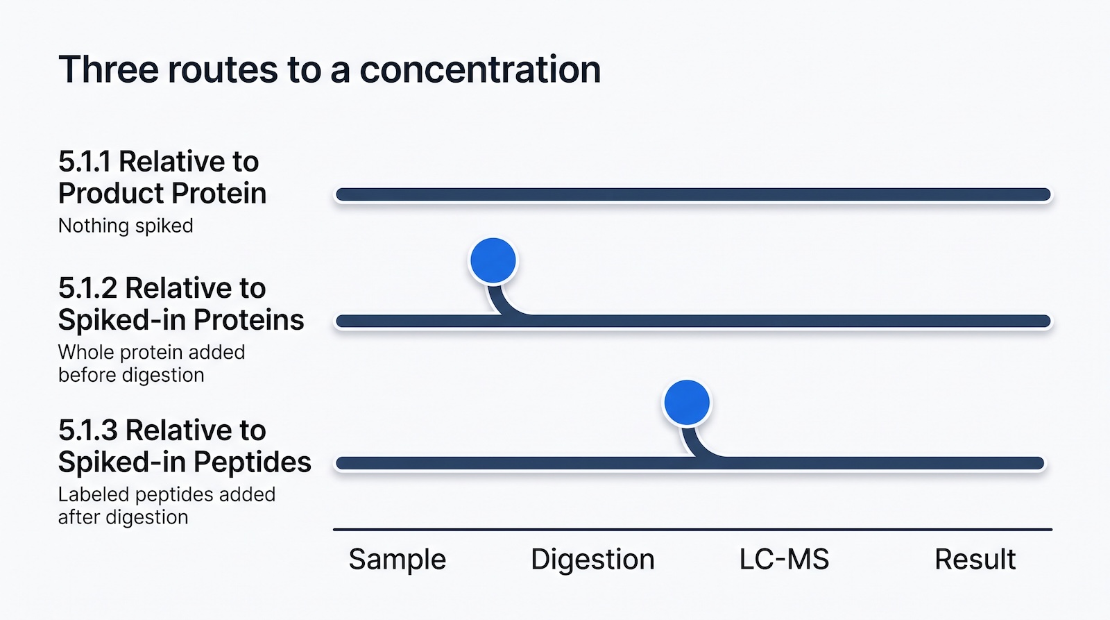
USP General Chapter <1132.1> describes three ways to put a number on a host cell protein measured by liquid chromatography-mass spectrometry, and it numbers them in Section 5.1: 5.1.1 Relative to Product Protein, 5.1.2 Relative to Spiked-in Proteins and 5.1.3 Relative to Spiked-in Peptides. Most of the industry calls them Method A, Method B and Method C. Those letters come from the qualification literature rather than from USP, and this page carries both so you can match what you read here to what you read in the chapter. The three are not interchangeable: they differ in what you add to the sample, what limits your quantitation range, and how far they carry you toward a qualified GMP method. This page covers each at the level of the bench, then what Section 5.2 asks of you for validation, what Section 5.3 asks for in system suitability, and what records a regulated lab needs.
Key facts
USP General Chapter <1132.1> is titled “Residual Host Cell Protein Measurement in Biopharmaceuticals by Liquid Chromatography-Mass Spectrometry”. It is informational, it became official on 1 May 2025, and its three quantitation methods are numbered, not lettered. The six points below are the ones a reader usually arrives looking for.
- USP General Chapter <1132.1> is titled “Residual Host Cell Protein Measurement in Biopharmaceuticals by Liquid Chromatography-Mass Spectrometry”.
- It is an informational chapter. USP states that General Chapters numbered 1000 to 1999 “are for informational purposes only. They contain no mandatory tests, assays, or other requirements applicable to any official article.”
- It was approved for publication on 1 November 2024 in USP-NF 2025 Issue 1 and became official on 1 May 2025. Four non-technical corrections announced on 31 January 2025 took effect on that same date.
- Section 5.1 sets out three quantitation methods and numbers them 5.1.1 Relative to Product Protein, 5.1.2 Relative to Spiked-in Proteins and 5.1.3 Relative to Spiked-in Peptides. The chapter never calls them Method A, B or C; those letters come from the qualification literature.
- Section 5.2 recognizes two routes. A product-specific method for release testing needs a matching standard or characterized reference material for every HCP quantified. More commonly, a qualified instrument and analysis system is used in relative quantitation mode, and that system may be considered to be in a state of validation separately from product-specific validation.
- USP catalogs stable isotope labeled heavy peptide analytical reference materials for nine named Chinese hamster ovary (CHO) host cell proteins, which removes the custom synthesis step from Section 5.1.3 for those targets.
What USP <1132.1> is, and what it is not
USP General Chapter <1132.1> is informational guidance on measuring residual host cell proteins by LC-MS. It is not a mandatory monograph requirement. USP’s own rule is that General Chapters numbered 1000 to 1999 “are for informational purposes only. They contain no mandatory tests, assays, or other requirements applicable to any official article” (USP, Identifying Official Text). Chapters below 1000 can become enforceable when a monograph or the General Notices reference them; <1132.1> sits above 1000, so it does not. It is the reference point you cite when you explain why your LC-MS host cell protein method looks the way it does, not a checklist you tick.
A host cell protein (HCP) is a process-related protein impurity produced by the expression system, such as a Chinese hamster ovary (CHO) cell line, and carried through purification alongside the product protein. HCPs are a critical quality attribute because of their potential effect on product quality, safety and efficacy. For background, see what a host cell protein is.
USP has set out its own reasoning in public. Anthony Blaszczyk and Niomi Peckham of USP’s Biologics group wrote up the chapter’s development for BioProcess Online in August 2023, recording that the proposed chapter “was published in March 2023 in the Pharmacopeial Forum (PF49(3)) for public comment” and stating USP’s technical position plainly: “Bottom-up LC-MS/MS is the standard HCP quantitation method” (Blaszczyk and Peckham, BioProcess Online, 18 August 2023). That is a USP-authored statement rather than a vendor claim, and it is the sentence to cite when someone asks why a bottom-up digest workflow is the default rather than an intact or top-down approach. The chapter was developed by the Biologics Monographs 2 (Proteins) Expert Committee.
On dates there is a persistent error in circulation. The chapter was approved for publication on 1 November 2024 in USP-NF 2025 Issue 1 (USP-NF notice), and that issue has a publication date of 1 November 2024 and an official date of 1 May 2025 (USP-NF Publication and Comment Schedule). Pages that give 1 November 2024 as the official date are quoting the publication date. Four corrections announced on 31 January 2025 took effect on 1 May 2025, including “product (or polysorbate)” becoming “product protein (or polysorbate)” in Section 4.1 (ECA Academy summary): a useful signpost to where the chapter handles those two interferences.


How the chapter is organized, and what it really calls the three methods
USP General Chapter <1132.1> does not use the labels Method A, Method B and Method C. It numbers its quantitation methods as subsections of Section 5.1 Methods for HCP Quantitation: 5.1.1 Relative to Product Protein, 5.1.2 Relative to Spiked-in Proteins and 5.1.3 Relative to Spiked-in Peptides. The letters are the convention of the qualification literature, above all of Chrone et al. (2025), the first published head-to-head qualification of all three, and they have since become the industry’s spoken shorthand.
Both are worth knowing, for different reasons. The letters are what a colleague, a vendor or a conference speaker will say, and they are what people type into a search box. The section numbers are what belongs in a method description, a development report or a response to a regulatory question. Naming Section 5.1.2 rather than Method B costs nothing and shows the reader you worked from the chapter rather than from a summary of it. This page leads with the section numbers and carries the letters alongside.
The chapter runs to eight numbered sections, followed by an acronym list and four references.
| Section | Title | What it covers |
|---|---|---|
| 1 | Introduction and Scope | Focus on recombinant proteins, with the general principles stated to be applicable to all types of biopharmaceuticals |
| 2 | Terminology | Definitions, including ng/mg and the synonyms in use for it |
| 3 | Introduction to Identification and Quantitation of HCPs Using LC-MS/MS Analysis | The bottom-up workflow in outline |
| 4 | Sample Preparation, Chromatographic Separation, and Mass Spectrometry Analysis | 4.1 Sample Preparation, 4.2 Chromatographic Methods, 4.3 Mass Spectrometry Analysis, 4.4 Data Analysis |
| 5 | Quantitation of HCPs | 5.1 Methods for HCP Quantitation (5.1.1, 5.1.2, 5.1.3), 5.2 Method Validation, 5.3 System Suitability |
| 6 | Reporting Results | Units, sensitivity statements and a multi-laboratory reproducibility case study |
| 7 | Best Practices for Reporting Data from ELISA Versus LC-MS/MS | 7.1 ELISA Quantitation Principles and Reporting Results, 7.2 Units of Measurement, 7.3 Comparison of ELISA and LC-MS/MS HCP Results, 7.4 Complementarity of LC-MS/MS and Total HCP ELISA |
| 8 | Discussion and Conclusions | Where each technique sits, and what would have to change for LC-MS/MS to take over release testing |
“Section numbers and titles of USP General Chapter <1132.1>, official 1 May 2025. Summarized in our own words; no chapter table is reproduced.”
The chapter’s own reference list has four entries and all four are worth reading: Huang L, Wang N, Mitchell CE, Brownlee T, Maple SR, De Felippis MR. “A novel sample preparation for shotgun proteomics characterization of HCPs in antibodies.” Analytical Chemistry 2017;89(10):5436-5444. Reference 1 of USP <1132.1>. https://doi.org/10.1021/acs.analchem.7b00304
USP <1132> versus USP <1132.1>: immunoassay versus LC-MS
USP <1132> covers immunoassay measurement of total residual HCP. USP <1132.1> covers LC-MS measurement of individual HCPs. They are companion chapters, not replacements. Most programs run both: an enzyme-linked immunosorbent assay (ELISA) for total HCP release testing, LC-MS for identity, risk assessment and orthogonal confirmation. There is a practical difference in access. All of <1132.1> sits behind a USP-NF subscription. For <1132>, USP posts the USP 39 and NF 34 version, official 1 May 2016, as a free full-text PDF. That free copy is worth reading first, because <1132.1> assumes its vocabulary. Note the version: it is the USP 39 and NF 34 text, and no Notice of Intent to Revise has been published for <1132> since it became official.
USP’s own guidance on where each chapter applies is a useful structural signal. In the USP mAb Analytical Guide, <1132> is cited at both the in-process testing stage and the release and stability stage, while <1132.1> appears only at release and stability. Read that as USP positioning LC-MS HCP measurement as a characterization and confirmation technique applied to finished material, rather than as an in-process monitoring tool. It is consistent with what <1132> itself says about orthogonal methods being applied “selectively on important lots”.
| Attribute | USP 1132 | USP 1132.1 |
|---|---|---|
| Full subject | Residual host cell protein measurement in biopharmaceuticals (immunoassay) | Residual host cell protein measurement in biopharmaceuticals by liquid chromatography-mass spectrometry |
| Core technique | Immunoassay, primarily ELISA | Bottom-up LC-MS/MS on digested samples |
| What it reports | Total HCP, as one number in ppm (ng HCP per mg product) | Individual HCPs, identified and quantified separately |
| Identity information | None. The assay cannot say which proteins responded | Protein-level identity from peptide sequence |
| Depends on | Anti-HCP antibody reagent and its coverage | FASTA database, digestion completeness and calibration standard |
| Typical role | Release and stability testing | Characterization, risk assessment, process development, orthogonal check on ELISA |
| Chapter status | Informational (numbered above 1000) | Informational (numbered above 1000) |
The two do not have to agree, and often do not. An ELISA reports only what its polyclonal antibody recognizes, which is why HCP antibody coverage is measured at all; LC-MS reports what the instrument detects and the software assigns. See why ELISA and LC-MS HCP numbers disagree.
Sections 5.1.1, 5.1.2 and 5.1.3 side by side
The three methods differ mainly in what plays the role of the calibration standard: the product protein in 5.1.1, a set of spiked intact proteins in 5.1.2, a synthetic peptide matched to a specific HCP peptide in 5.1.3. Everything else, digestion, chromatography, acquisition, follows from that choice.
| Attribute | 5.1.1 Relative to Product Protein (Method A) | 5.1.2 Relative to Spiked-in Proteins (Method B) | 5.1.3 Relative to Spiked-in Peptides (Method C) |
|---|---|---|---|
| What you add to the sample | Nothing | One or more intact proteins at known concentration | Synthetic peptides at known concentration, usually stable isotope labeled (SIL) |
| When you add it | Not applicable | Before enzymatic digestion | Before or after digestion |
| What you calibrate against | Peptides from the product protein at a known product concentration | Summed peptide signal from the spiked proteins, converted to a response factor | The SIL partner of each target HCP peptide |
| Core assumption | Product protein peptides and HCP peptides ionize and fragment comparably, and both sit inside the linear range | The spiked proteins digest with the same efficiency as the HCPs and give a representative median response factor | Digestion of the target HCP goes to completion, and the SIL peptide behaves identically to its native counterpart |
| Demonstrated linear range of the calibrant (Chrone et al. 2025) | 117,647 to 1,000,000 ng/mL, R-squared 0.991 | 10 to 15,000 ng/mL, R-squared 0.998 | 0.4 to 205 ng/mL, R-squared 1.000 |
| What sets the upper limit | The product protein level | The amount of spiked-in protein (ng protein/mL) | The spiked peptide level relative to the target peptide (ng peptide/mL) |
| Reagent cost | None | Low. Commodity proteins | High for a new target, since each peptide is custom synthesized. Moderate for the nine CHO HCPs USP catalogs as heavy peptide analytical reference materials |
| Setup effort | Lowest. No spike step, no new reagents | Moderate. One spike step, one qualification of the standard mix | Highest. Peptide selection, synthesis, purity and stability control, and MRM method development if targeted |
| Untargeted or targeted | Untargeted | Both, using the same standard proteins | Best in targeted mode. Usable untargeted with care |
| Outcome in the published qualification | Acceptable linearity and high precision, conditional accuracy, could not meet the pre-defined specificity criteria | Met all criteria | Met all criteria, with interference on one peptide in untargeted mode |
| Principal limitation the chapter names | MS1 intrascan dynamic range. A highly purified product protein is typically more than four orders of magnitude above the HCPs, while many Orbitrap instruments cover two to three orders within a scan | The standards have to be well characterized for purity and concentration, must yield unique peptides, and must not create ambiguous or false-positive HCP identifications | Cost, because ideally every HCP would have its own synthetic SIL peptide, and the assumption that digestion is 100% efficient with complete peptide recovery |
| When a lab chooses it | Early discovery and process development screening, where relative change matters more than an absolute number | Development through to QC and GMP, when you want one method that covers unknown and known HCPs | Release-grade measurement of named high-risk HCPs where sensitivity and specificity must be defensible |
The linear range row comes from Chrone et al. (2025), the first published head-to-head qualification of all three methods, run on a purified monoclonal antibody spiked with CHO harvested cell culture fluid, measuring clusterin and lipoprotein lipase (J Pharm Biomed Anal 265:117051). Those are working ranges of the calibration standard, not of the HCP.
Section 5.1.1 Relative to Product Protein (Method A)
Section 5.1.1 uses peptides from the product protein itself as the calibration standard. You digest the sample, measure the signal from a defined set of product protein peptides and from a defined set of HCP peptides, then scale by the known product concentration and the molecular weight ratio. Nothing is spiked in. As Chrone et al. put it, “peptides from the product protein can be used as a standard for relative quantitation of HCPs.”
It costs nothing and works on any sample you have. The limitation is the concentration gap, and the chapter states it as an instrument specification rather than a general worry. A highly purified biopharmaceutical protein is typically more than four orders of magnitude more abundant than the HCPs around it, while many Orbitrap instruments deliver only two to three orders of magnitude of intrascan dynamic range at MS1. Quantitation relative to the product protein therefore may not suit MS1-level work at all, which is a concrete reason to prefer a spiked standard rather than a matter of taste. In practice the product protein’s high-intensity peptides sit above the upper limit of quantitation, which is why Chrone et al. advise against picking product peptides the way you would pick Hi3 peptides. You have to hunt for moderate-intensity product peptides that stay linear, and that selection is a bias you will have to defend. For how Hi3 works when the target is the HCP, see Hi3 peptide quantification.
Section 5.1.1 also has a structural problem with specificity. Demonstrating specificity means comparing a sample with and without the standard, and you cannot remove the product protein without changing the matrix, so the published study could not complete a specificity assessment for it. A mock process run with a non-expressing cell line, or affinity depletion of the product, “could theoretically enable specificity testing” in the authors’ words, but neither is trivial.
Their verdict: Section 5.1.1 “demonstrated acceptable linearity, high precision (CVs < 20%), and a conditional demonstration of accuracy. However, it could not meet the pre-defined criteria for specificity in this study”, and “is best suited for untargeted early discovery stages.” Use it for screening process changes, where direction matters more than a defensible absolute figure. Do not build a release method on it.
Section 5.1.2 Relative to Spiked-in Proteins (Method B)
Section 5.1.2 adds one or more intact proteins at known concentration before digestion, then uses their summed peptide signal to derive a response factor that converts HCP peptide signal into mass. Because the standards go in before digestion, they experience the same reduction, alkylation, protease exposure, desalting and injection as the HCPs, so the response factor normalizes for digest variability.
The chapter sets four requirements on the standards and one on the arithmetic. Each standard protein must be well characterized for purity and supplied at a known concentration; it must generate peptides unique to itself; it must not create ambiguous or false-positive HCP identifications in the search; and the response factor used for the sample is the median across the set of standards rather than any single one. The published qualification spiked seven standard proteins at 2,000 ng/mL: myoglobin, beta-lactoglobulin, lysozyme C, hemoglobin subunit alpha, hemoglobin subunit beta, alpha-lactalbumin and serotransferrin. Taking the median across the seven protects against one standard behaving badly, and summing all observed peptides returns HCP mass without needing a theoretical molecular weight for the full-length sequence. Chrone et al. warn that “caution is needed when using protein standards from the same or homologous species”: a bovine or human standard can share tryptic peptides with a CHO HCP and inflate the reading.
Section 5.1.2 is what most labs should default to. It covers known and unknown HCPs with the same reagents, works in untargeted and targeted acquisition, and in the published study it “showed high linearity (R² = 1), a quantitative range of 2-4 orders of magnitude, and high precision (CVs < 20%)”. The authors judged it “suitable for the entire product development process, and potential method validation for QC and GMP analysis.” The cost is one pipetting step and one qualification of the standard mix.
Section 5.1.3 Relative to Spiked-in Peptides (Method C)
Section 5.1.3 adds synthetic peptides at known concentration, matched in sequence to specific peptides of specific HCPs, and quantifies each HCP from the signal ratio between the native peptide and its spiked partner. The peptides are usually stable isotope-labeled (SIL), meaning they carry heavy carbon-13 or nitrogen-15 so they co-elute with the native peptide but separate by mass.
This is the most sensitive and most specific of the three, and the most work. The chapter is candid about why. Ideally every HCP you report would have its own synthetic SIL peptide, and the reason labs do not do that is cost. It advises choosing at least three or four peptide standards per target, because in practice some turn out to be unsuitable through interference or insufficient sensitivity, so you need spares before you commit. And the whole approach assumes digestion efficiency of 100% with complete recovery of the peptide, which is the assumption most likely to be wrong. You must also know which HCPs you are measuring before you start, have the peptides synthesized, and control their purity and stability as reagents. The published study used six SIL peptides, three for clusterin and three for lipoprotein lipase, spiked at 63 ng/mL.
Beyond sensitivity, the advantage is identification confidence: a SIL peptide has the same retention time and fragment ion pattern as its native counterpart, so a matching pair is strong evidence the assignment is correct. That matters most for high-risk host cell proteins where a false positive or a missed detection has consequences, such as lipoprotein lipase and clusterin.
Two cautions follow from that. The digestion assumption bites because you are comparing a synthetic intact peptide against one that had to be released from a protein, so any shortfall in digestion is read as a shortfall in HCP. And different peptides from the same protein give different answers: three SIL peptides for clusterin returned 171, 930 and 422 ng/mL, a 5.4-fold spread, while precision within each peptide stayed high. The effect is not confined to one study. Kreimer et al. (2017), one of the chapter’s own four references, compared quantitation against protein standards with quantitation against SIL peptide standards and found the two approaches can differ by two to three fold, attributing the gap to peptide ionization efficiency, protein digestion efficiency and similar factors (Anal Chem 89(10):5294-5302). Read that as the working accuracy envelope when you move between 5.1.2 and 5.1.3 on the same sample. Fix your peptide, fix your method, and report the number against that specific calibration standard rather than as absolute truth. The practicality objection to Section 5.1.3 is weaker than it was. The standard argument against it is that custom peptide synthesis, purity control and stability control make it too slow and too expensive for anything but a flagship target. That was true when every heavy peptide had to be commissioned. USP now catalogs stable isotope labeled heavy peptide analytical reference materials covering nine named CHO host cell proteins, supplied in 50 µg units, so for those targets you buy a characterized reagent with a catalog number instead of commissioning one.
| CHO host cell protein | USP catalog number | Notes |
|---|---|---|
| Cathepsin D | 1130704 | Heavy Peptide 1 |
| Glutathione-S-transferase P1 | 1130711 | Heavy Peptide 1 |
| Peroxiredoxin 1 | 1130725 | Heavy Peptide 3 |
| Protein disulfide isomerase (PDI) | 1130727 | |
| Clusterin | 1130730, 1130741, 1130752 | Three heavy peptides |
| Lipoprotein lipase (LPL) | 1130763, 1130774, 1130785 | Three heavy peptides |
| Phospholipase B-like 2 (PLBL2) | 1130796 |
USP also supplies purified recombinant CHO PLBL2 protein (1582716), usable as a Section 5.1.2 standard for that target, and CHO Null Cell Harvest Cell Culture Fluid (1544913) as a spiking matrix (USP monoclonal antibody standards and analytical reference materials list). Read the category label carefully before writing any of this into a protocol: USP states that analytical reference materials “are different from USP Reference Standards and not required for compendial compliance”. They are characterized materials you can cite and buy, not compendial standards that confer compliance by themselves.
That changes the verdict in the method table. Section 5.1.3 is still the highest-effort option, and peptide selection, interference checking and MRM development remain real work. But for the nine proteins above, and for clusterin and lipoprotein lipase in particular, the synthesis step drops out, and those are the targets most often driving a release-grade individual HCP specification. If your risk assessment names one of those nine, Section 5.1.3 is a more reasonable proposition than its reputation suggests. See also the USP HCP SIL peptide application note.

Sample preparation the chapter asks you to think about
Sample preparation is where most LC-MS HCP methods succeed or fail, because the product protein is in vast excess and formulation components interfere. Section 4.1 addresses removal of the product protein or polysorbate, HCP enrichment, and native and denatured digestion protocols, and it tabulates the caveats attached to each option. One of those options is immunoaffinity enrichment using the immobilized polyclonal anti-HCP antibodies from your ELISA, which can also be turned around to assess the coverage of those reagents by comparing the HCPs identified in the eluate with those identified in the load. The chapter attaches an important caveat to it: the approach can only capture HCPs the polyclonal mixture holds antibodies against, and it is not orthogonal to the ELISA it borrows the reagents from. Four decisions matter in practice.
Product protein handling. Depleting the product, for example by affinity capture, raises the relative abundance of HCPs and improves detection at low ppm. It also changes the matrix, complicating accuracy and specificity claims, and can co-deplete HCPs that bind the product. Native digestion, where the folded product resists protease while HCPs are cleaved, is the alternative.
Polysorbate. Polysorbate 20 and polysorbate 80 are surfactants used to stabilize protein formulations. They suppress ionization and contaminate the source, so formulated drug product usually needs the polysorbate removed or the sample diluted before injection. Note the circularity: lipoprotein lipase is an HCP you want measured precisely because it degrades polysorbate.
Digestion. Reduction, alkylation and proteolysis must be reproducible before any of the three methods gives a stable number. The published study used dithiothreitol at 6.9 mM for 10 minutes at 85 degrees Celsius, iodoacetamide at 16.6 mM for 45 minutes at room temperature, then sequential lysyl endopeptidase and trypsin, both at 0.01 micrograms per microliter for 60 minutes at 37 degrees Celsius, on a pipetting robot. Digestion variability propagates straight into the result, and Methods B and C correct for it only if the standard is spiked before digestion.
Acquisition mode. Data-independent acquisition (DIA) fragments every precursor in defined mass windows, so the data is complete and can be re-interrogated later. Data-dependent acquisition (DDA) selects precursors on the fly and is biased toward abundant species. Multiple reaction monitoring (MRM) is a targeted mode watching defined transitions. The published study used DIA for untargeted work and MRM for targeted Section 5.1.3 work, and the targeted mode resolved the one specificity failure DIA showed. See DIA versus DDA.
Do you have to validate it? Section 5.2 gives two different answers
Section 5.2 Method Validation separates two situations, and most laboratories are in the second one without realizing the chapter allows it.
Product-specific method validation, for release testing. If you are measuring an individual HCP to release a drug substance, the chapter expects the procedure to be validated in line with ICH guidance, and it expects a matching standard or a stable, characterized reference material, a stable isotope labeled peptide included, for every HCP you quantify. That is a real burden: one qualified standard per analyte, each with its own purity, concentration and stability control, plus the full validation package on the product.
A qualified system used in relative quantitation mode. The chapter describes the more common route as qualifying the instrument and the analysis system with reference materials, using them to evaluate system suitability, and then running the method in relative quantitation mode. A system qualified that way may be considered to be “in a state of validation” (USP <1132.1>, Section 5.2) separately from product-specific drug substance method validation.
That distinction is the practical answer to “do I have to validate this”, and it is the part of the chapter most often skipped over. It means untargeted profiling, clearance studies, process characterization and orthogonal confirmation of an ELISA do not each demand a validated release method. It also relocates the work. The burden moves off the product and onto the system: the reference materials you qualify with, the system suitability sample described below, the data analysis settings you fix in advance, and the records that show all three were in place on the day the sample ran. That is a software and records problem as much as an instrument problem.
Where a product-specific validation is required, ICH Q2(R2) is the framework the chapter points you at.
Qualifying an HCP LC-MS method against ICH Q2(R2)
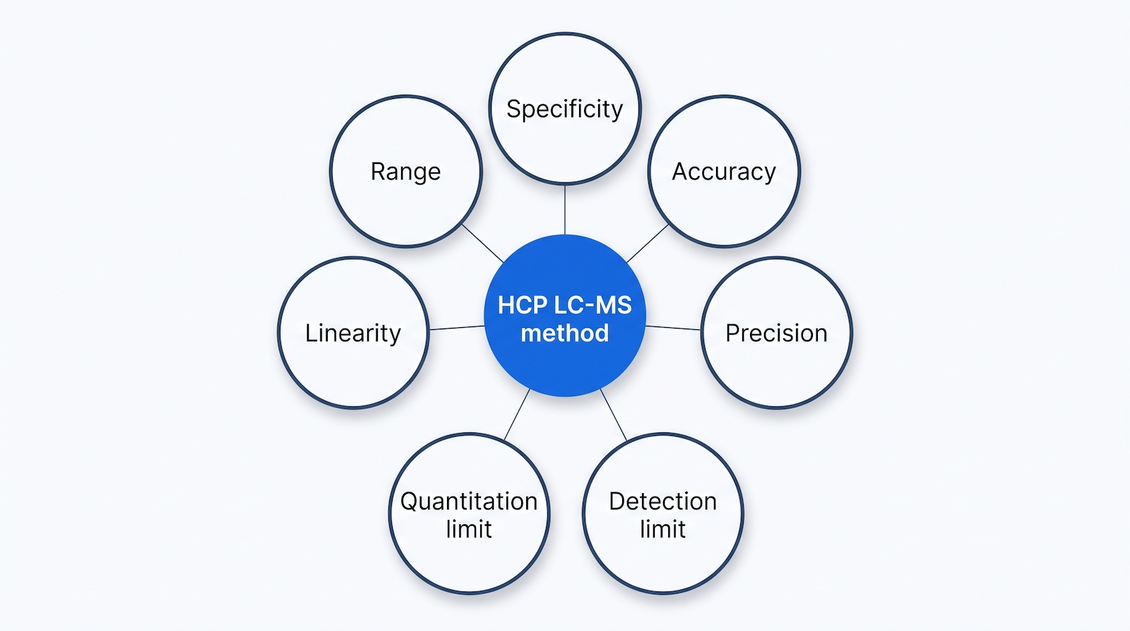
ICH Q2(R2) “Validation of Analytical Procedures” reached Step 4 on 1 November 2023 and sets out the characteristics you demonstrate: specificity and selectivity, range, response and linearity, accuracy, precision, detection limit, quantitation limit and robustness (ICH Q2(R2) guideline PDF; FDA guidance page). An LC-MS HCP method is unusual because the analyte is a moving population of proteins rather than one molecule, so you qualify against named representative HCPs and argue from there.
| ICH Q2(R2) characteristic | What you actually do in an HCP LC-MS qualification |
|---|---|
| Specificity and selectivity | Compare samples with and without the spiked standard and test whether the HCP result shifts. Chrone et al. used a two-tailed t-test with P more than 0.05 as the criterion for no significant difference. Confirm identity by peptide sequence, retention time and, for Section 5.1.3, co-elution with the SIL partner. Section 5.1.1 cannot be tested this way because the product cannot be removed without altering the matrix. |
| Range | State the range over which the calibration standard is linear and accurate, and confirm it brackets your expected HCP levels. The range is set by the product protein in Section 5.1.1, by the spiked protein amount in Section 5.1.2, and by the spiked peptide amount in Section 5.1.3. |
| Response and linearity | Build a dilution series of the calibration standard across at least five consecutive levels. Chrone et al. used R-squared greater than or equal to 0.980 across five or more points as the criterion, and reported 0.991, 0.998 and 1.000 for Sections 5.1.1, 5.1.2 and 5.1.3. |
| Accuracy | Spike a known amount of the standard, or of a characterized HCP material, and calculate recovery. Chrone et al. accepted 50 to 200 percent recovery at a representative spike level, which is wider than a small-molecule assay and reflects the reality of protein quantitation by MS. |
| Precision, repeatability | Replicate preparations of the same sample by one analyst on one instrument on one day. Report coefficient of variation per HCP and per peptide. |
| Precision, intermediate | Vary analyst, day and instrument. Chrone et al. used triplicate preparations across three days, two instruments and two analysts, at least nine determinations, with a criterion of CV less than or equal to 30 percent. All three methods came in under 20 percent. |
| Detection limit and quantitation limit | Determine the lowest level that still meets your accuracy and precision criteria, per HCP and per peptide, not per method in the abstract. |
| Robustness | Vary digestion time, protease lot, column lot, gradient and injection volume deliberately, and show the result holds. This is where automation of sample preparation pays for itself. |
| System suitability | Run before and within every analytical sequence, built around a spiked HCP at a known level. See below. |
Section 5.3 System Suitability: the spike that protects a negative result
System suitability is the check you run at the start of a sequence, and at intervals within it, to prove the instrument and method are performing well enough for the results to count. Section 5.3 recommends a system suitability sample, and it is specific about what that sample should be: an HCP spiked into a sample of the protein product at a known level, with detection of that spike serving as the benchmark for the sensitivity of the run.
The reason the chapter gives for it is the useful part. The spike matters most when no HCPs are identified, or when the result is not the one you expected. A negative finding is only worth as much as the evidence that the measurement was working when it was made, and without a spike, “none detected” and “nothing worked” produce the same report. If you take one thing from Section 5.3 into your SOP, take that: the clean result is the one that needs the spike.
Build the rest of the check around that spiked HCP. Four groups of criteria cover it in practice.
- The spiked system suitability injection. Inject the product sample spiked with a known level of a known HCP, and confirm it is detected and quantified within a pre-set window. A reference digest or a commercial peptide retention time standard can sit alongside it, but it does not replace it.
- Chromatographic criteria. Retention time reproducibility within a stated window, peak width at half height below a stated value, and no carryover above a stated threshold in a blank following the highest standard.
- Mass spectrometric criteria. Mass accuracy within a stated ppm window, signal intensity for named peptides above a floor, and for MRM the expected transition ratios.
- Quantitative criteria. The response factor, or the light-to-heavy ratio for a SIL peptide at a known level, inside a pre-set range. This is the check that protects the number you report.
Record pass or fail against pre-defined limits and define the action on failure before you start. Where the run reports no HCPs at all, the system suitability record is the evidence, so it belongs in the report rather than in an instrument log nobody reads. Our guide to validated proteomics software covers the difference between research tools and tools you can qualify.
Note: Section 5.3 recommends the spiked system suitability sample and the sensitivity benchmark. The four groups of chromatographic, mass spectrometric and quantitative criteria below the spike are practice-based, not prescribed by the chapter, and are offered as a worked example.

Implementing this in a regulated lab: the sequence
- Define the question. Total HCP for release, or named individual HCPs for risk assessment? For release, USP <1132> and an ELISA remain the primary route with LC-MS orthogonal. For individual HCPs, continue.
- List the HCPs that matter. Build a short list from process knowledge, published CHO risk lists and your own untargeted data. Clusterin and lipoprotein lipase are on almost every list.
- Pick the method. Section 5.1.1 for early screening, Section 5.1.2 as the default from development to GMP, Section 5.1.3 for named high-risk HCPs needing the lowest limits. Many labs run 5.1.2 and 5.1.3 together: the spiked proteins give the profile, the spiked peptides give the defensible number on the proteins that matter.
- Source and qualify the standard. For Section 5.1.2, characterize the standard proteins for purity and concentration and check in silico that their tryptic peptides neither collide with CHO HCP peptides nor create false-positive identifications. For Section 5.1.3, select at least three or four peptides per HCP because some will prove unsuitable, source SIL versions, and qualify purity and stability.
- Fix the sample preparation. Product depletion or native digestion, polysorbate handling, digestion protocol. Automate it and write it down as a method, not a note.
- Fix the acquisition method. Column, gradient, acquisition mode and, for targeted work, the transition list. Freeze it before qualification starts.
- Fix the data analysis settings. FASTA database and version, false discovery rate, peptide confidence threshold, minimum peptides per protein, extraction window, and the rule for which peptides feed quantitation. Chrone et al. used a 1 percent false discovery rate, a 95 percent peptide confidence threshold, a minimum of two peptides per identification and a 75 ppm extraction window, and flagged any CV above 0.3 for manual review.
- Decide which validation route you are on. A product-specific method for release testing goes through the full ICH Q2(R2) table above with a characterized standard per HCP. A qualified instrument and analysis system running in relative quantitation mode is the other route Section 5.2 recognizes, and it is the right one for profiling, clearance and orthogonal work. Write down which one you chose and why, before generating data.
- Write the method and the report. State the chapter section (5.1.1, 5.1.2 or 5.1.3), the calibration standard and its lot, the demonstrated range and the units. Report as nanograms of HCP per milligram of product protein, a unit the chapter’s Terminology also records as having parts per million as a common synonym, and say which section and standard the figure is relative to, because the answer moves with the calibrant. How HCP ppm is calculated sets out the arithmetic.
- Close the records loop. A GMP lab needs an audit trail of every analysis and reanalysis, electronic signatures on review and approval, user access control, version control on raw data and analysis, and archived raw files that cannot be silently altered. That is the 21 CFR Part 11 and EU Annex 11 layer: see our 21 CFR Part 11 compliance guide, how to implement 21 CFR Part 11 and ALCOA and ALCOA+.
The software layer, and why it needs checking
Whichever method you pick, the number you report comes out of a database search engine, and Section 4.4 Data Analysis is blunt about that. Search engines were developed for proteomics, HCP work pushes them down to low-abundance peptides with poor signal to noise, and the chapter tells the user to expect some false positives and warns that “search engine output should not be trusted implicitly” (USP <1132.1>, Section 4.4), especially for low-abundance HCPs. It treats manual inspection of peptide-level raw data, the extracted ion chromatogram trace, the MS spectrum and the MS/MS spectrum, as a routine part of HCP data analysis rather than as an escalation, and it is most insistent about that for low-abundance HCPs and drug substance samples. That is not a criticism of any search engine. It is a statement about signal-to-noise at the bottom of a five or six order of magnitude dynamic range, where one misassigned peptide can create or erase an HCP. The practical consequences are the ones in step 7: constrain the search, require more than one peptide per identification (two unique peptides is the chapter’s description of common practice, with extra confirmatory work expected where an identification rests on one), control the false discovery rate, and make peptide-level evidence visible rather than buried. In a regulated setting, add that every setting is recorded, versioned and attributable, so a result can be reconstructed two years later.
Where TotalLab fits
SpotMap MS is TotalLab’s LC-MS host cell protein analysis software. It compares LC-MS traces against a user-supplied FASTA file using data-independent acquisition (DIA), is designed to support alignment of LC-MS HCP workflows with USP <1132.1>, and pairs with the AuditSafe overlay for 21 CFR Part 11 and EU Annex 11 audit trails, electronic signatures and user permissions. For evaluation criteria see USP <1132.1> HCP software; for make-or-buy, see bringing HCP analysis in-house.
See the SpotMap MS product page, start a free trial, or talk to us about qualifying a Section 5.1.2 or 5.1.3 workflow in your lab.
Frequently asked questions
Is USP <1132.1> mandatory?
No. USP General Chapter <1132.1> is an informational chapter. USP states that chapters numbered 1000 to 1999 are for informational purposes only and contain no mandatory tests, assays or other requirements applicable to any official article. It is guidance you are expected to have considered when you justify your host cell protein control strategy, not a requirement you can be cited against directly.
When did USP <1132.1> become official?
It was approved for publication on 1 November 2024 in USP-NF 2025 Issue 1, and USP-NF 2025 Issue 1 became official on 1 May 2025. Four minor, non-technical corrections were announced on 31 January 2025 and took effect on the same date. Sources that give 1 November 2024 as the official date are quoting the publication date.
Does USP <1132.1> call its quantitation methods A, B and C?
No. The chapter numbers them as subsections of Section 5.1: 5.1.1 Relative to Product Protein, 5.1.2 Relative to Spiked-in Proteins and 5.1.3 Relative to Spiked-in Peptides. The letters A, B and C are the shorthand used in the qualification literature, particularly Chrone et al. (2025), and they have spread from there into everyday use. Both refer to the same three methods. Use the section numbers in any document a reviewer will read.
Which method should I use: 5.1.1, 5.1.2 or 5.1.3?
Section 5.1.2, relative to spiked-in proteins, is the sensible default for most labs. It covers known and unknown host cell proteins with one set of reagents, normalizes for digestion variability, and in the published qualification it met every ICH Q2(R2) criterion tested. Use 5.1.1 only for early screening, and add 5.1.3 for named high-risk HCPs where you need the lowest quantitation limits.
Do I need SIL peptides?
Only for Section 5.1.3. Stable isotope labeled peptides give the best sensitivity and the strongest identity evidence, because they co-elute with the native peptide and share its fragmentation pattern. The chapter advises picking at least three or four candidate peptides per target because some prove unsuitable, and notes the approach assumes complete digestion. USP now sells CHO HCP SIL peptide standards, including clusterin and lipoprotein lipase.
Do I have to validate an LC-MS HCP method?
It depends which route you are on. Section 5.2 expects a product-specific method used for release testing of individual HCPs to be validated per ICH guidance, with a matching standard or characterized reference material for every HCP quantified. More commonly the instrument and analysis system are qualified with reference materials, evaluated for system suitability and used in relative quantitation mode, and that qualified system may be considered to be in a state of validation separately from product-specific validation.
Why do two SIL peptides for the same HCP give different answers?
Because Section 5.1.3 measures a peptide, not a protein, and then infers the protein. Digestion efficiency, peptide recovery and ionization differ between peptides. In the published qualification, three SIL peptides for clusterin returned 171, 930 and 422 ng/mL, a 5.4-fold spread. Kreimer et al. (2017), a reference the chapter itself cites, found protein standards and SIL peptide standards can differ two to three fold.
What does system suitability look like for an HCP LC-MS run?
Section 5.3 recommends a system suitability sample consisting of an HCP spiked into the protein product at a known level, with detection of that spike as the sensitivity benchmark. The chapter stresses that this matters most when nothing is found or the result is unexpected, because a negative result needs evidence that the measurement was working. Add chromatographic, mass accuracy and quantitative criteria around it, with pre-defined limits.
References
- United States Pharmacopeial Convention. General Chapter <1132.1> Residual Host Cell Protein Measurement in Biopharmaceuticals by Liquid Chromatography-Mass Spectrometry (notice of publication). 2024. https://www.uspnf.com/notices/gc-1132-1-nitr-20241101
- United States Pharmacopeial Convention. General Chapter <1132.1> Residual Host Cell Protein Measurement in Biopharmaceuticals by Liquid Chromatography-Mass Spectrometry. USP-NF, official 1 May 2025. DOI 10.31003/USPNF_M17756_03_01. Subscription access. Sections cited on this page: 1 (Introduction and Scope), 4.1 (Sample Preparation), 4.4 (Data Analysis), 5.1.1, 5.1.2 and 5.1.3 (Methods for HCP Quantitation), 5.2 (Method Validation), 5.3 (System Suitability) and 6 (Reporting Results). https://doi.usp.org/USPNF/USPNF_M17756_02_01.html
- United States Pharmacopeial Convention. Identifying Official Text (FAQ on mandatory versus informational general chapters). https://www.usp.org/frequently-asked-questions/identifying-official-text
- United States Pharmacopeial Convention. USP-NF Publication and Comment Schedule. https://www.uspnf.com/print/pdf/node/16296
- ECA Academy. USP Announces Corrections to General Chapter <1132.1> on Residual Host Cell Protein Measurement. 2025. https://www.gmp-compliance.org/gmp-news/usp-announces-corrections-to-general-chapter-1132-1-on-residual-host-cell-protein-measurement
- Chrone, V.G., Blaszczyk, A.J., Zhang, D.H., Nielsen, S.B., Crawford, J., Schwämmle, V., Højrup, P., Kofoed, T., Peckham, N., Mørtz, E. Host cell protein quantitation by LC-MS. Experimental demonstration, qualification, and comparison of methods in USP 1132.1. Journal of Pharmaceutical and Biomedical Analysis, vol. 265, article 117051, 15 November 2025. DOI 10.1016/j.jpba.2025.117051. PMID 40639094. https://www.sciencedirect.com/science/article/pii/S0731708525003929 (open access PDF: https://alphalyse.com/wp-content/uploads/2025/07/victor-chrone-et-al-host-cell-protein-quantitation-by-lc-ms-experimental-demonstration.pdf)
- International Council for Harmonisation. ICH Harmonised Guideline Q2(R2): Validation of Analytical Procedures. Step 4, 1 November 2023. https://database.ich.org/sites/default/files/ICH_Q2%28R2%29_Guideline_2023_1130.pdf
- U.S. Food and Drug Administration. Q2(R2) Validation of Analytical Procedures (guidance for industry, final, March 2024). https://www.fda.gov/regulatory-information/search-fda-guidance-documents/q2r2-validation-analytical-procedures
- United States Pharmacopeial Convention. Quantitation of CHO Host Cell Proteins Using Stable Isotope Labeled (SIL) Peptides (application note BIO.1023.A). https://www.usp.org/sites/default/files/usp/document/our-work/biologics/app-notes/BIO.1023.A.HCPSILPeptide.ApplicationNote.pdf
- BioPharm International. Quantifying Host Cell Proteins by LC-MS: Experimental Demonstration and Method Comparison in USP 1132.1. 2025. https://www.biopharminternational.com/view/quantifying-host-cell-proteins-by-lc-ms-experimental-demonstration-and-method-comparison-in-usp-1132-1
- Alphalyse. USP 1132.1: How to perform HCP analysis by MS. https://alphalyse.com/usp-chapter-1132-1/
- United States Pharmacopeial Convention. Monoclonal Antibody (mAb)-related USP-NF Standards and Analytical Reference Materials. 7 October 2025. Source for the nine CHO HCP heavy peptide catalog numbers, the recombinant PLBL2 protein and the CHO null cell HCCF material. https://www.usp.org/sites/default/files/usp-bio_monoclonalantibodies-standards-materials_07oct2025.pdf
- Blaszczyk, A., Peckham, N. (United States Pharmacopeia). USP Unpacks The Evolving HCP Identification And Quantitation Story Behind <1132.1>. BioProcess Online, 18 August 2023. https://www.bioprocessonline.com/doc/usp-unpacks-the-evolving-hcp-identification-and-quantitation-story-behind-0001
- United States Pharmacopeial Convention. General Chapter <1132> Residual Host Cell Protein Measurement in Biopharmaceuticals. USP 39 and NF 34, official 1 May 2016. The companion immunoassay chapter to <1132.1>. Free full-text PDF posted by USP. Note that this free copy is the 2016 version and that no Notice of Intent to Revise has been published for <1132> since it became official. https://www.usp.org/sites/default/files/usp/document/our-work/biologics/USPNF810G-GC-1132-2017-01.pdf
- United States Pharmacopeial Convention. mAb Analytical Guide: In-Process Testing. https://www.usp.org/mab-analytical-guide/in-process-testing
- United States Pharmacopeial Convention. mAb Analytical Guide: Release and Stability. https://www.usp.org/mab-analytical-guide/release-and-stability
- Kreimer, S., Gao, Y., Ray, S., Jin, M., Tan, Z., Mussa, N.A., Tao, L., Li, Z., Ivanov, A.R., Karger, B.L. Host Cell Protein Profiling by Targeted and Untargeted Analysis of Data Independent Acquisition Mass Spectrometry Data with Parallel Reaction Monitoring Verification. Analytical Chemistry, 2017, 89(10):5294-5302. Cited by USP <1132.1> as its own reference 4; source for the two to three fold difference between protein standards and SIL peptide standards. DOI 10.1021/acs.analchem.6b04892. https://doi.org/10.1021/acs.analchem.6b04892
- Huang L, Wang N, Mitchell CE, Brownlee T, Maple SR, De Felippis MR. “A novel sample preparation for shotgun proteomics characterization of HCPs in antibodies.” Analytical Chemistry 2017;89(10):5436-5444. Reference 1 of USP <1132.1>. https://doi.org/10.1021/acs.analchem.7b00304
- Krey JF, Wilmarth PA, Shin JB, Klimek J, Sherman NE, Jeffery ED, Choi D, David LL, Barr-Gillespie PG. “Accurate label-free protein quantitation with high- and low-resolution mass spectrometers.” Journal of Proteome Research 2014;13(2):1034-1044. Reference 3 of USP <1132.1>. https://doi.org/10.1021/pr401017h