Guide
The plaque assay in viral vector development
How it works, what the pharmacopoeias require, and how to get a titer in 24 hours

Guide
How it works, what the pharmacopoeias require, and how to get a titer in 24 hours
If your product is a virus, infectivity is the attribute that matters most. A vector that cannot enter a cell cannot deliver its payload, and an oncolytic virus that cannot replicate cannot kill a tumor. That is why every regulator asks for an infectious titer, and why the plaque assay, first used in animal virology in the 1950s, is still on the release and stability panels of oncolytic virus, viral vaccine and many viral vector programs today.
It is also slow. Depending on the virus, plaques take from 3 to 14 days to form, and the count at the end is done by eye. This guide explains how the plaque assay works, where it sits in pharmaceutical quality control, what the European Pharmacopoeia, EMA and FDA say about infectious titer, and how a kinetic imaging approach now delivers a validated-quality titer 24 hours after infection.
On this page
The plaque assay counts infectious virus particles. You infect a layer of susceptible cells with a diluted sample, then hold the virus in place with a thick overlay so each infectious particle can only spread to the cells next to it. Over the following days, each original infection grows into a small zone of dead or damaged cells. That zone is a plaque.
Because one plaque starts from one infectious unit, counting plaques and correcting for the dilution gives you plaque forming units per milliliter (PFU/mL). That number is a functional titer. It tells you how much of your material can actually infect and replicate, which a physical measurement cannot.
This is the distinction that makes the assay matter. Genome titers by qPCR or ddPCR, particle counts by nanoparticle tracking and capsid ELISAs are fast, but none of them can tell an infectious virion from a damaged one. As the analytical team behind the kinetic titer method put it in 2024, “The use of cell-based assays is therefore the gold standard to determine infectious virus titers.” [1]
The method varies by virus and cell line, but the core workflow is the same in most labs.
Add the time to expand and seed cells, and a single plaque titer routinely takes a working week or more from start to result. Repeat determinations for precision, and the week becomes two.
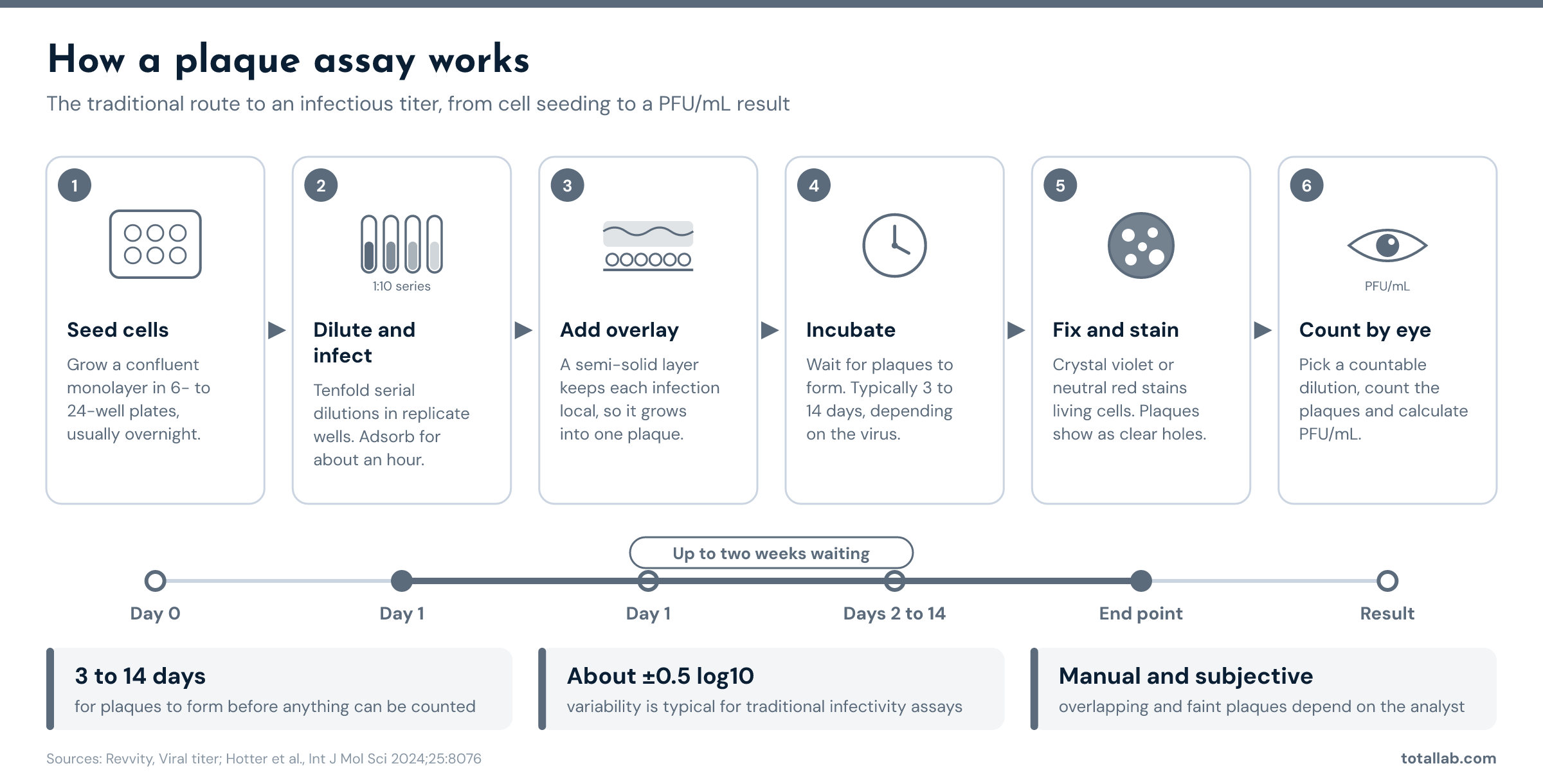
The traditional plaque assay. Most of the time to result is spent waiting for plaques to form.
For most virus-based medicines, infectious titer is the critical quality attribute. It reflects potency, and for many products it is the number that defines the dose given to the patient. [1]
That puts the infectivity assay in three places in a GMP program.
Upstream of release, the same assay drives process development and formulation work, where hundreds of samples need a titer each before a decision can be made. This is where a slow assay costs the most.
The pharmacopoeias and guidelines are clear that infectious titer must be measured. They are equally clear that the plaque assay is one acceptable way to measure it, not the only one.
European Pharmacopoeia. In March 2024 the European Pharmacopoeia Commission adopted a new general monograph, Gene therapy medicinal products for human use (3186), with an accompanying general chapter, Additional information on gene therapy medicinal products for human use (5.34). Together they replace general chapter 5.14, Gene transfer medicinal products for human use, which had stood since 2006. The new texts were published in Supplement 11.7 and became mandatory on 1 April 2025. Monograph 3186 covers the product classes already approved in Europe, including adeno-associated virus vectors and oncolytic herpes simplex virus. Chapter 5.34 carries the revised sections for adenovirus, poxvirus and retroviridae-derived vectors. [2] [3]
The way the Ph. Eur. words infectivity tests is worth noting. Its 2024 chapter on phage therapy medicinal products (5.31) states that the infectious phage titre is “determined by a plaque assay or any other suitable method.” [4] The pharmacopoeia names the classical method and leaves room for a validated alternative. The same principle runs through the gene therapy texts, which ask for a titration in cell culture against a reference standard.
EMA. The EMA guideline on the quality, non-clinical and clinical aspects of gene therapy medicinal products sets the expectation in plain terms: “For viral vectors, infectious titre should be quantified”. It also asks that the number of particles be determined and that the particle to infectivity ratio be included to define the content of the drug substance. [5]
FDA. The FDA’s January 2020 guidance, Chemistry, Manufacturing, and Control (CMC) Information for Human Gene Therapy Investigational New Drug Applications (INDs), asks sponsors to provide the CMC information needed to assure the product’s safety, identity, quality, purity and strength, including potency. For a replicating or infectious viral product, an infectious titer is a core part of that package. [6]
USP. USP general chapter <1237> Virology Test Methods describes the virology test methods used in developing biological products. [7]
ICH. Whatever method you choose, it has to be validated for its intended use. ICH Q2(R2), Validation of Analytical Procedures, sets out what that means for accuracy, precision, specificity and range. [8]
The practical reading for a QC lab is this. You must report a validated infectious titer for release and stability. The plaque assay is the reference method most people know, but the texts allow any suitable, validated cell-based method.
The plaque assay has lasted because it is cheap, needs no special equipment and works for a wide range of virus and cell combinations. Its weaknesses show up when you scale it.
None of this makes the plaque assay wrong. It makes it the bottleneck.
The TCID50 assay is the other classical infectivity method. Instead of counting plaques, it scores replicate wells at each dilution as infected or not, then uses the Spearman-Kärber or Reed-Muench method to estimate the dilution that infects half the wells. It avoids the overlay and suits viruses that do not form clean plaques, but it still waits for a late endpoint and still uses a dilution series per sample.
The kinetic infectious virus titer (KIT) method takes a different approach. It images living cells every hour after infection, measures the early change in cell shape that infection causes, and fits a curve to the whole time course instead of reading a single endpoint. The table compares the three for a typical batch of 24 samples.
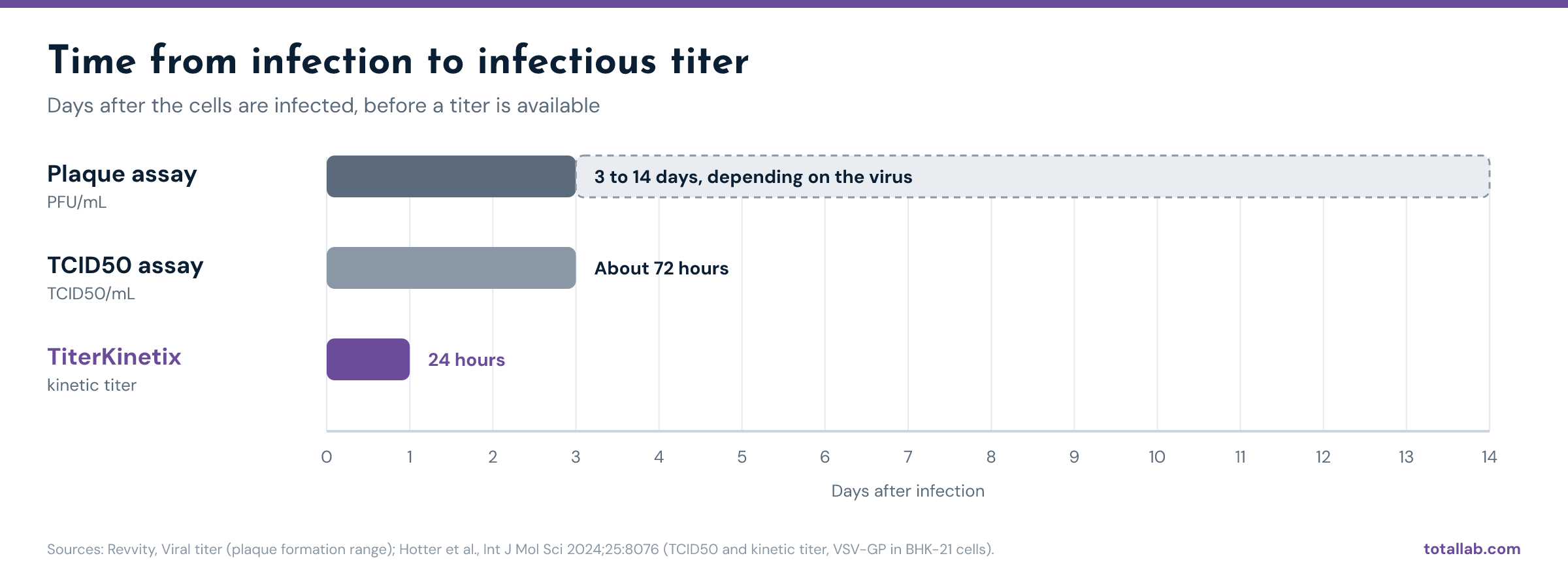
Time from infection to result. TCID50 and kinetic figures are from Hotter et al. 2024; the plaque range is across viruses.
| Parameter | Plaque assay | TCID50 assay | Kinetic titer (TiterKinetix) |
|---|---|---|---|
| What is measured | Discrete plaques under an overlay | Infected or uninfected wells across a dilution series | Infection-induced cell rounding over time |
| Unit reported | PFU/mL | TCID50/mL | Same unit as the reference standard (PFU/mL or TCID50/mL) |
| Time from infection to result | 3 to 14 days depending on virus | About 72 hours in the published comparison | 24 hours |
| Overlay or stain needed | Yes | Optional stain | No |
| Readout | Manual plaque count | Microscopic or image-based CPE scoring | Automated kinetic image analysis |
| Wells per sample | Full dilution series in replicate | 8 replicates per half-log dilution | 3 wells at a single dose |
| Plate format | 6- to 24-well plates | One 96-well plate per sample | Up to 24 samples on one 96-well plate |
| Precision | About ±0.5 log10 is typical | About 44% CV for one determination | 11% intra-plate and 17% intermediate CV |
| Stability-indicating | Yes | Yes | Yes |
| Works for non-cytopathic vectors such as AAV | No | Yes with qPCR or ddPCR readout | No |
Plaque assay, TCID50 and kinetic titer compared. Kinetic and TCID50 figures are from Hotter et al. 2024, which compared the two methods for VSV-GP in BHK-21 cells. Plaque formation time is a published range across viruses. Your numbers will depend on your virus and cell line.
The 24-hour figure comes from published science. In 2024, analytical scientists at a global pharmaceutical company’s Viral Therapeutics Center published the kinetic infectious virus titer (KIT) assay in the International Journal of Molecular Sciences. [1] They found that infected cells round up in a time- and dose-dependent way within hours of infection, well before plaques or full cytopathic effect appear. Tracking that rounding in label-free bright-field images, and fitting a curve to every time point, gave them an infectious titer within 24 hours of infection.
The published results set the benchmark.
TiterKinetix is TotalLab’s software implementation of that method. It reads the time-lapse images your plate imager already captures, on an Agilent Cytation 5, a Sartorius Incucyte or another automated imager running 96- or 384-well plates. It then classifies cells in every image, fits the kinetic curves, builds the standard curve from your reference standard and interpolates each sample’s titer, with QC metrics and a report. There is no overlay, no staining step and no manual count.
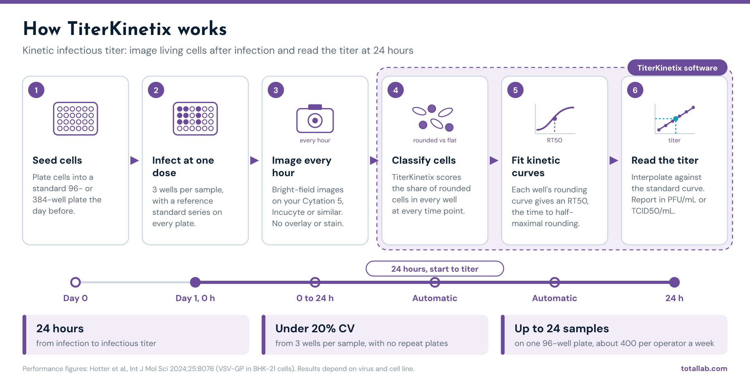
The kinetic titer workflow. Steps 4 to 6 run automatically in TiterKinetix, and the titer is ready 24 hours after infection.
Two features matter in particular for a release lab.
It reports in your existing units. KIT is a relative method. Titers are read against a reference standard on every plate, and they come out in whatever unit that standard was characterized in. If your reference standard carries a plaque assay value, TiterKinetix reports PFU/mL. The 2024 paper did exactly this for parapoxvirus ovis, defining the dose in PFU per cell. [1] That gives you a direct bridge from the plaque assay you validated to a faster method, and a clean comparability package for your regulator.
It is built for regulated use. TiterKinetix runs with TotalLab’s AuditSafe module, which provides audit trails, electronic signatures, granular user permissions and image authenticity verification for FDA 21 CFR Part 11 and EU Annex 11. TotalLab has been building life science software in Newcastle since 1989, and regulated 21 CFR Part 11 software since 2009.
A note on validation. Moving a release assay from plaque assay to a kinetic method is a method change. You will validate the KIT assay for your product under ICH Q2(R2) and file the change with your regulator. The pharmacopoeial texts allow a suitable alternative method. TiterKinetix gives you the precision and audit trail that make the validation package strong.
Kinetic titer works for viruses that cause a measurable change in cell shape soon after infection. The 2024 paper demonstrated it for three unrelated viruses: the oncolytic VSV-GP in BHK-21 cells, Newcastle disease virus in DF-1 chicken fibroblasts, and parapoxvirus ovis in HeLa cells. [1] The authors expect it to extend to many other viruses that cause cytopathic effect, with some tuning of cell density, MOI range and curve model for each virus and cell combination. Candidates include other VSV-based products, adenovirus vectors, oncolytic herpes simplex virus and poxvirus vectors.
It is not a fit for everything, and it is better to say so here.
If your virus causes cytopathic effect and your samples are in the working range, kinetic titer can take days out of every release and stability test.
It measures the infectious titer of a virus product in plaque forming units per milliliter. In pharma it supports release and stability testing of oncolytic viruses, live viral vaccines and some viral vectors, and it is used to characterize virus seed stocks and reference standards.
Plaque formation alone takes 3 to 14 days depending on the virus. With cell seeding, staining and counting, a single result commonly takes a week or more.
No. Ph. Eur. general monograph 3186 and chapter 5.34, the EMA gene therapy quality guideline and FDA’s 2020 CMC guidance require an infectious titer, measured by a suitable and validated method. The Ph. Eur. phage therapy chapter says it outright: a plaque assay or any other suitable method.
A plaque assay counts discrete zones of infection under an overlay and reports PFU/mL. A TCID50 assay scores whole wells as infected or not across a dilution series and estimates the dose that infects 50% of wells. TCID50 values are usually higher than PFU values for the same sample, so the two units are not interchangeable without a conversion study. [10]
Yes, for cytopathic viruses. The kinetic infectious virus titer method reads infection-induced cell rounding from hourly images and gives a titer 24 hours after infection, with under 20% CV from three wells per sample. TiterKinetix automates that analysis.
Yes. Titers are calculated against your reference standard and reported in the same unit. If your reference standard was characterized by plaque assay, results are in PFU/mL.
See TiterKinetix run on your own virus, cell line and imager. We will show you how kinetic titers compare with your plaque assay or TCID50 data and what a bridging study would look like.
Working from endpoint data today? Try the free TCID50 calculator.