Guide
Precipitin test
How it works, the main types and how to measure the results

Guide
How it works, the main types and how to measure the results
A precipitin test detects an antigen or an antibody by the visible precipitate the two form when they meet. Soluble antigen and antibody bind into a cross-linked lattice, and when their amounts are in balance the lattice grows large enough to fall out of solution as a white ring, line or halo. Where and how much precipitate forms tells you whether the antigen or antibody is present, whether two antigens are related, or, in radial immunodiffusion, how much antigen there is.
This guide covers how the reaction works, every common format from the ring test to radial immunodiffusion, where the test is still used in forensic science, vaccine testing and clinical diagnosis, and how to measure the result from an image instead of a ruler.
On this page
| Test | How it is set up | What you read | Result | Typical use |
|---|---|---|---|---|
| Ring (tube) test | Antigen layered over antiserum in a narrow tube | A white ring at the interface | Qualitative | Quick presence test; forensic species identification |
| Ouchterlony double immunodiffusion | Antigen and antibody in separate wells cut in a gel | Precipitin lines between the wells and how they join or cross | Qualitative | Comparing antigens; antibody specificity; serum precipitins |
| Radial immunodiffusion (RID or SRID) | Antibody mixed into the gel; antigen in a well | The diameter or area of the ring around each well | Quantitative | Antigen concentration; influenza vaccine potency |
| Immunoelectrophoresis | Proteins separated by electrophoresis then antiserum diffused from a trough | Position and shape of each precipitin arc | Qualitative | Identifying proteins in serum |
| Rocket immunoelectrophoresis | Antigen driven by electrophoresis into antibody-containing gel | The height of each rocket-shaped precipitate | Quantitative | Fast antigen measurement |
| Counter-current immunoelectrophoresis (CIEP) | Antigen and antibody driven toward each other from opposite wells | A precipitin line between the wells | Qualitative | Forensic species tests on stains; serum precipitins |
The main precipitin test formats, what you read from each, and what each is used for.
A precipitin is an antibody that forms a precipitate with its soluble antigen. The antigen that triggers it is sometimes called the precipitinogen. The word is also used loosely for the precipitate itself, the white antigen-antibody complex you see in the tube or gel.
Precipitation needs a soluble antigen with several binding sites (epitopes) and antibodies that can bridge between antigen molecules. Polyclonal antisera work best because their mixture of antibodies binds many different epitopes and builds a large lattice. A single monoclonal antibody that recognizes one epitope often cannot cross-link enough to precipitate.
Precipitation and agglutination are the same idea at different scales. In precipitation the antigen is a soluble molecule, such as a serum protein. In agglutination the antigen sits on a particle, such as a red blood cell or a bacterium, and the antibody clumps the particles together.
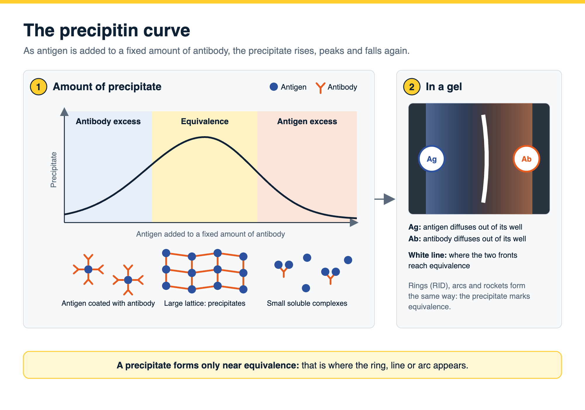
In the 1930s, Michael Heidelberger and Forrest Kendall measured precipitation chemically: they added increasing amounts of antigen to a fixed amount of antiserum and measured the nitrogen in each precipitate. The amount of precipitate rose, peaked and fell again, and they derived equations that described the whole curve (ref 1). That curve has three zones:
Every precipitin test is a way of finding the zone of equivalence. In a gel, antigen and antibody diffuse toward each other from opposite sides and form concentration gradients, so equivalence is reached somewhere along the way, and that is exactly where the line, arc or ring appears.
The ring test is the simplest format. Antiserum is placed in a narrow tube and the antigen solution is layered gently on top, so the two liquids meet at a sharp interface without mixing. As they diffuse into each other, a white ring of precipitate forms at the interface if the antigen and antibody match.
A positive result is a white band at the junction between the two layers. No band means no reaction at the concentrations tested, so dilutions of the antigen are often run side by side. The ring test is quick and needs no gel, but it is qualitative and it is easily spoiled by mixing the layers or by cloudy samples.
In 1949 Örjan Ouchterlony described antigen-antibody reactions in gels (ref 2). In the Ouchterlony test, wells are cut in an agar or agarose plate, antibody goes in one well, usually the center, and antigens go in the wells around it. Both diffuse outward through the gel, and a precipitin line forms wherever they meet at equivalence.
The shape of the lines between two neighboring antigen wells compares the antigens:
Ouchterlony plates are used to check antibody specificity, compare antigens and, as below, detect serum precipitins in patients. The result is qualitative: the number, position and shape of the lines.
Radial immunodiffusion (RID, or single radial immunodiffusion, SRID) makes the precipitin reaction quantitative. Antibody is mixed evenly into the gel, and antigen is placed in a well. As the antigen diffuses outward it precipitates with the antibody in a ring, and the more antigen in the well, the further it travels before reaching equivalence, so the larger the ring.
There are two ways to read it:
Either way, standards of known concentration are run on the same plate and the unknowns are read off the standard curve. Fahey and McKelvey used it to measure serum immunoglobulins (ref 4). It is still the accepted potency assay for inactivated influenza vaccines: SRID, which measures the vaccine’s hemagglutinin content, has been the standard potency method since 1978 (refs 5, 6), and national control laboratories and manufacturers check their SRID proficiency against each other (ref 7).
Adding an electric field makes precipitin tests faster or more informative:
The best-known use of the precipitin test is to answer one forensic question: is this blood, or tissue, human? An extract of the stain is tested against antiserum raised to human serum proteins. A precipitin line or ring with anti-human serum, and none with antisera to other species, identifies the stain as human. The same antisera panel identifies animal species.
The method works on material other than fresh blood. Researchers identified the species of tooth fragments stored for up to six months in dry or humid conditions by counter-current electrophoresis (ref 11), and forensic serology methods have been used to identify tissue, bone and bloodstains recovered at aircraft accident sites (ref 12). In soil from an ambush scene in Kosovo, sampled about a year and a half after the event, crossover immunoelectrophoresis detected human blood proteins in 44 of 72 samples (61%) (ref 13).
DNA analysis has largely replaced immunological tests in forensic laboratories, but the same study concluded that in particular situations the precipitin approach may still be a valuable tool (ref 13).
“Serum precipitins” are antibodies in a patient’s blood that precipitate with an environmental or fungal antigen. They are detected with double immunodiffusion or counter-current electrophoresis against a panel of antigens.
How you read a precipitin test depends on the format:
Two things decide whether the numbers are any good. First, the image: photograph or scan the plate evenly lit against a dark background, with a scale in the frame or a calibrated camera, and keep the image as an uncompressed TIFF. Faint lines are easier to see after the gel is washed and stained with a protein stain. Second, the measurement: a ring read with a ruler or calipers depends on where each person decides the edge is, and that variation goes straight into the standard curve. Measuring every ring with the same edge setting removes it.
Phoretix Toolbox is TotalLab’s general-purpose image measurement software, for experiments that do not fit a dedicated gel, blot or plate workflow (ref 19). For precipitin plates it replaces the ruler:
Phoretix Toolbox reads 8-bit and 16-bit TIFF images as well as JPEG, PNG, BMP and IMG, and runs on Windows, macOS and Linux (ref 19). For regulated work such as SRID potency testing of vaccine lots, it can be used with AuditSafe, which adds secure sign-in, audit trails, electronic signatures and image authenticity checks for 21 CFR Part 11 (ref 19).

Where the precipitin lines run along a strip, as on an immunodiffusion or immunoelectrophoresis strip, Phoretix 1D can read them as bands in a lane profile instead, giving the position and volume of each line, as in the image above.
The video below shows the Phoretix Toolbox auto-trace tool: setting the threshold, completing a shape, reading the area, height and width, reusing the same setting on the next object and exporting the results.

Open an image of your own plate in a free Phoretix Toolbox trial: trace every ring or rocket with one threshold and export the measurements for your standard curve.
A: A test that detects an antigen or an antibody by the visible precipitate they form together. When soluble antigen and antibody meet in balanced amounts they cross-link into a lattice that falls out of solution as a white ring, line or halo.
A: To identify the species a blood, tissue, bone or tooth sample came from. An extract is tested against antisera to human and animal proteins, and a precipitate with anti-human serum identifies it as human. DNA analysis has largely replaced it, but it still has a place in particular situations.
A: At the zone of equivalence, where antigen and antibody are in balance. In a ring test that is the interface between the antiserum and the antigen solution layered on top; in a gel it is the point between the wells where the two diffusing fronts meet.
A: The ratio of antigen to antibody at which they cross-link into the largest lattice and precipitation is at its maximum. With excess antibody, the antigen runs out and less precipitate forms; with excess antigen, the complexes stay small and soluble and the precipitate dissolves.
A: The soluble antigen that stimulates the production of precipitins and reacts with them to form a precipitate.
A: In Ouchterlony double immunodiffusion, both antigen and antibody diffuse from wells and the pattern of lines compares antigens qualitatively. In radial immunodiffusion, antibody is spread through the gel and only antigen diffuses, so the size of the ring measures how much antigen there is.
A: Measure the diameter, or the area, of each ring and compare it with standards of known concentration on the same plate. With the endpoint method the area is proportional to concentration; with the kinetic method the diameter is proportional to its logarithm. Measuring every ring with the same edge setting in image analysis software removes reader-to-reader variation.
Your software won't do what you need? Ask TotalLab →Tell us the problem; we reply within two working days.